Как правильно пишется слово «лимфоузел»
лимфоу́зел
лимфоу́зел, -узла́
Источник: Орфографический
академический ресурс «Академос» Института русского языка им. В.В. Виноградова РАН (словарная база
2020)
Делаем Карту слов лучше вместе
Привет! Меня зовут Лампобот, я компьютерная программа, которая помогает делать
Карту слов. Я отлично
умею считать, но пока плохо понимаю, как устроен ваш мир. Помоги мне разобраться!
Спасибо! Я стал чуточку лучше понимать мир эмоций.
Вопрос: символизированный — это что-то нейтральное, положительное или отрицательное?
Синонимы к слову «лимфоузел»
Предложения со словом «лимфоузел»
- После наших операций мы дружно ходили откачивать скапливающуюся лимфу из места, где был удалён лимфоузел.
- Лимфоузел является биологическим фильтром, в котором образуются лимфоциты – живые защитники организма, уничтожающие чужеродные клетки и болезнетворные бактерии.
- Всего-навсего воспалился лимфоузел – врачи назвали это лимфоденитом, выписали кучу таблеток и запретили беспокоиться.
- (все предложения)
Отправить комментарий
Дополнительно
А Б В Г Д Е Ж З И Й К Л М Н О П Р С Т У Ф Х Ц Ч Ш Щ Э Ю Я
лимфоу́зел, -узла́
Рядом по алфавиту:
лимфогранулемато́зный
лимфодрена́ж , -а, тв. -ем
лимфодрена́жный
лимфо́зный
лимфолейко́з , -а
лимфологи́ческий
лимфоло́гия , -и
лимфо́ма , -ы
лимфообраще́ние , -я
лимфоотто́к , -а
лимфоретикулёз , -а
лимфосарко́ма , -ы
лимфосисте́ма , -ы
лимфото́к , -а
лимфотро́пный
лимфоу́зел , -узла́
лимфоцита́рный
лимфоци́тный
лимфоцито́з , -а
лимфоци́ты , -ов, ед. -ци́т, -а
ли́мцы , -ев, ед. ли́мец, ли́мца, тв. ли́мцем (от Ли́ма)
линалоо́л , -а
лина́рия , -и
линга́ла , нескл., м. и неизм.
лингафо́н , -а
лингафо́нный
лингая́ты , -ов, ед. -я́т, -а (секта)
ли́нгва фра́нка , нескл., м.
лингва́льный
лингватулёз , -а
лингвату́лы , -ов, ед. -ту́л, -а
Что такое лимфоузлы? Исчерпывающий ответ на заданный вопрос вы найдете в материалах статьи. Кроме этого, мы расскажем о строении представленного органа, а также о причинах его воспаления, возможных последствиях и так далее.
Общие сведения
Что такое лимфоузлы? Лимфатическим узлом называют периферический орган лимфатической системы, который выполняет функцию природного фильтра. Через него протекает вся лимфа, поступающая от различных частей и органов тела. В организме человека выделяют несколько групп таких узлов, которые называют регионарными.
Размер лимфоузлов
Внешне лимфатические узлы выглядят как округлые, овальные, бобовидные или иногда лентовидные образования. Их размеры варьируются в пределах 0,5-50 миллиметров и более. Как известно, такие периферические органы окрашены в серовато-розовый цвет. Лимфатические узлы находятся только по ходу лимфатических сосудов и располагаются гроздьями до десяти штук возле крупных вен и кровеносных сосудов.
Внешний вид
Лимфоузлы человека покрыты соединительнотканной оболочкой, от которой внутрь органа отходят так называемые трабекулы или балки. Они представляют собой своеобразные опорные структуры. Следует особо отметить, что сам периферический орган, выполняющий функцию природного фильтра, состоит из стромы. Она образовывается из ретикулярной соединительной ткани, на которой имеются отростчатые клетки, формирующие трехмерную сеть. Помимо этого, строма состоит из фагоцитирующих веществ (или макрофагов), представленных в лимфоузлах несколькими разновидностями.
Внутреннее строение лимфоузла
На разрезе лимфатических узлов сразу же выделяются две главные зоны. Ближе к оболочке – корковое вещество. В нем различают поверхностную часть и область глубокой коры (или так называемый паракортикальный слой). К внутренней зоне лимфатического узла относят мозговое вещество.
Все пространство данного органа заполнено лимфоидной тканью. В зоне поверхностной коры, которая находится ближе к оболочке, располагаются небольшие узелки или фолликулы. Следует отметить, что они имеют центральную светлую часть (герминативный центр), где происходит дифференцировка B-лимфоцитов и антигензависимая пролиферация, а также темную поверхностную, которая содержит в себе большое количество плотно расположенных друг к другу и довольно мелких лимфоцитов.
Принцип работы
В паракортикальной зоне лимфоциты располагаются равномерно и очень плотно. В этой части органа преобладают T-лимфоциты. Здесь они проходят антигензависимую дифференцировку и пролиферацию. Что же касается мозгового вещества, то скопления лимфоидной ткани в нем представлены мозговыми тяжами (или мякотными шнурами), куда из поверхностной коры мигрируют B-лимфоциты.
Принцип работы этого периферического органа заключается в следующем: лимфа притекает к узлам по подходящим с выпуклой стороны сосудам, а оттекает по выносящим с вогнутой части. При этом внутри узла лимфа довольно медленно просачивается по пространствам, называемыми синусами. Они располагаются между оболочкой и трабекулами, а также лимфоидной тканью.
Так же, как и сосуды, внутреннее пространство узла имеет собственную выстилку, которая образуется литоральными или береговыми клетками. Как правило, их отростки направляются внутрь синуса, где они начинают контактировать с ретикулярными клетками. Следует особо отметить, что в отличие от обычных сосудов у синусов нет свободной полости, ведь она полностью перегорожена трехмерной сетью. За счет такого строения лимфа, попадая в узел, медленно просачивается, что способствует ее тщательному очищению от инородных тел. Также данный процесс происходит и благодаря макрофагам, располагающимся по самому краю лимфоидных скоплений. Кстати, во время прохождения по синусам (мозговому веществу) лимфа в полной мере насыщается и антителами, продуцирующими плазматическими клетками тяжу (мозговую).
Для чего нужны лимфатические узлы?
Что такое лимфоузлы, мы выяснили. Сейчас же хочется рассказать о том, для чего вообще нужны эти органы. Дело в том, что протекающая лимфа приносит в узел так называемые чужеродные антигены. В результате это приводит к развитию в органах реакций иммунного ответа. В зависимости от вида и характера инородных тел подобные реакции могут активно развиваться во внешних или внутренних зонах. Это приводит к едва заметному или сильному увеличению размеров узлов. Таким образом, можно смело отметить, что представленные периферические органы являются своеобразным барьером для распространения не только различных инфекций, но и раковой опухоли. Ведь в узле способны созревать защитные клетки, которые принимают активное участие в уничтожении чужеродных антигенов и других веществ.
Где располагаются лимфатические узлы?
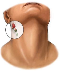
Лимфоузлы (фото представлены в данной статье) находятся в теле человека достаточно большими группами, которых насчитывается около десяти штук. Располагаются они так, чтобы препятствовать развитию различных инфекций и раковой опухоли. Именно по этой причине узлы находятся возле самых важных для жизни органов и систем, а именно в локтевых и коленных сгибах, в подмышечных впадинах и паховой области. Кроме этого, они располагаются в области шеи, грудной и брюшной полости. Таким образом, лимфатические узлы обеспечивают полную защиту от различных инфекций и опухолей головы.
Виды лимфоузлов
Следует особо отметить, что такая система фильтрации находится не только в вышеуказанных местах. Лимфокапилляры пронизывают и все внутренние органы. При этом они выполняют те же самые функции.
Итак, в теле человека имеется несколько групп лимфатических узлов, а именно:
- внутригрудные;
- бронхопульмональные;
- локтевые;
- селезеночные;
- парааортальные;
- брыжеечные;
- подвздошные (внешние, внутренние, а также общие);
- паховые (поверхностные и глубокие);
- бедренные;
- подколенные.
Почему увеличиваются лимфатические узлы?
Причины увеличения лимфоузлов – самые разные заболевания. При этом следует особо отметить, что появившаяся шишка свидетельствует о неблагополучии именно той зоны, в которой она находится. Чаще всего увеличение лимфоузлов связано с какими-либо инфекциями. Кроме того, такая патология возникает на фоне опухолевого поражения.
Итак, давайте рассмотрим более подробно, почему и при каких заболеваниях происходит увеличение лимфоузлов у детей и взрослых:
- Гнойные процессы. Как правило, при таком отклонении возникает так называемый острый лимфаденит. Чаще всего это происходит в результате попадания микробов из ран, которые расположены в зоне нахождения того или иного периферического органа. К основным симптомам данного воспаления можно отнести появление болезненности при пальпациях и покраснение кожи. Если в такой момент не вскрыть образовавшуюся шишку, то оболочка узла порвется, а гной проникнет в окружающие его ткани, в результате чего возникнет довольно тяжелое осложнение, называемое флегмоном.
- Увеличенные лимфоузлы у детей довольно часто свидетельствуют о наличии туберкулеза. Как правило, при таком заболевании шишки образуются в грудной полости и на шее.
- Нередко причиной увеличения лимфатических узлов у маленьких детей является микроб бартонелла. Переносчиками такой бактерии выступают кошки, чьи царапины можно довольно часто наблюдать у ребенка. Именно через эти ранки микроб очень быстро распространяется по лимфососудам и попадает в узлы, которые впоследствии увеличиваются и становятся довольно болезненными. Таким образом, долго не заживающая гнойная рана, а также шишка, появившаяся рядом, всегда должна наводить на мысль о развитии болезни «кошачьей царапины».
- При ОРВИ у взрослых и детей может быть вплоть до нескольких групп увеличенных лимфоузлов. Причиной такого отклонения является избыточный ответ иммунной системы на вторжение в организм больного каких-либо вирусов. Следует заметить, что лимфоузлы в таких ситуациях увеличиваются не очень сильно, но при ощупывании они довольно болезненны.
- Венерические заболевания, в частности, сифилис, тоже становятся причиной увеличенных лимфатических узлов. При этом больной может наблюдать у себя шишки в паховой области, а также язвочки на половых органах. В отличие от других заболеваний при сифилисе увеличенные лимфоузлы могут быть безболезненными, а соответственно, и незаметными для человека.
- Длительно не проходящие увеличенные группы лимфатических узлов могут свидетельствовать о наличии таких серьезных заболеваний, как листериоз, бруцеллез, ВИЧ-инфекция или мононуклеоз.
Увеличение узлов при опухолях
Опухоль узлов может возникнуть из-за лимфопролиферативных заболеваний (если изначально опухоль произошла от лимфоузла), а также от метастатического поражения. К первому отклонению, прежде всего, относится лимфосаркома и лимфогранулематоз. Лимфатические узлы при таких заболеваниях увеличиваются до четырех-пяти сантиметров и становятся довольно плотными. Однако при ощупывании образовавшиеся шишки не являются болезненными. Кстати, при первоначальном увеличении внутрибрюшных или внутригрудных лимфоузлов такие заболевания могут быть и не распознаны.
Подведем итоги
Теперь вам известно, что такое лимфоузлы. Следует особо отметить, что увеличение органов периферической системы должно сразу же насторожить больного. Причина этого проста: такое патологическое состояние свидетельствует о том, что в организме человека происходят опасные для его жизни и здоровья процессы. В этом случае рекомендуется сразу же обратиться к врачу и пройти полное медицинское обследование.
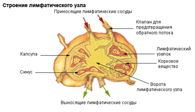
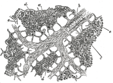
Структура лимфатического узла.
Лимфати́ческий у́зел (лимфоузел) — периферический орган лимфатической системы, выполняющий функцию биологического фильтра, через который протекает лимфа, поступающая от органов и частей тела.
В теле человека выделяют около 150 групп лимфоузлов, называемых регионарными.
Анатомия и физиология
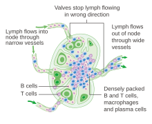
Структура лимфатического узла и течение лимфы через лимфатические синусы.
Лимфатические узлы представляют собой образования округлой, овальной, бобовидной, реже лентовидной формы размерами от 0,5 до 50 мм и более. Лимфоузлы окрашены в розовато-серый цвет. Лимфатические узлы располагаются по ходу лимфатических сосудов, как правило, гроздьями до десяти штук, возле кровеносных сосудов, чаще — возле крупных вен.
Поверхность лимфатического узла покрыта соединительнотканной капсулой, от которой внутрь узла отходят трабекулы — балки, также образованные соединительной тканью. Они представляют собой опорные структуры. Строма — основа лимфатического узла образована ретикулярной соединительной тканью, отростчатые клетки которой и, образованные ими ретикулярные волокна, формируют трехмерную сеть. В состав стромы входят также фагоцитирующие клетки — макрофаги, представленные в лимфатических узлах несколькими разновидностями.
На разрезе органа выделяются две основные зоны. Ближе к капсуле — корковое вещество, в котором различают поверхностную часть и зону глубокой коры (паракортикальную зону). Внутренняя часть лимфатического узла получила название мозговое вещество.
Внутреннее пространство органа содержит скопления лимфоидной ткани. В области поверхностной коры, ближе к капсуле располагаются лимфатические узелки (фолликулы). На окрашенных препаратах они имеют более светлую центральную часть — герминативный центр, в котором происходит антигензависимая пролиферация и дифференцировка B-лимфоцитов (бурсазависимая зона). Поверхностная, более темная на препаратах часть узелка — лимфоидная корона содержит большое количество мелких, плотно расположенных лимфоцитов.
В зоне глубокой коры (паракортикальной зоне) лимфоциты располагаются плотно, довольно равномерно. В этой области преобладают T-лимфоциты, которые проходят здесь антигензависимую пролиферацию и дифференцировку (тимусзависимая зона).
В мозговом веществе скопления лимфоидной ткани представлены мозговыми тяжами (мякотными шнурами), в которые мигрируют B-лимфоциты из поверхностной коры. B-лимфоциты дифференцируются окончательно в плазматические клетки, продуцирующие иммуноглобулины — антитела.
Лимфа притекает к лимфатическим узлам по приносящим лимфатическим сосудам, подходящим к узлу с выпуклой стороны, и оттекает по выносящему лимфатическому сосуду, отходящему с вогнутой стороны узла в области ворот. Внутри узла лимфа медлено протекает (просачивается) по внутренним пространствам, которые называются лимфатическими синусами. Синусы располагаются между капсулой, трабекулами и скоплениями лимфоидной ткани. Как и сосуды, синусы имеют собственную выстилку, образованную литоральными (береговыми) клетками. Их отростки направлены внутрь синуса, где они контактируют с отросками ретикулярных клеток. Таким образом, в отличие от сосудов синусы не имеют свободной полости, она перегорожена трехмерной сетью, образованой ретикулярными и литоральными клетками, благодаря этому лимфа медлено просачивается по синусам. Это способствует её очищению от инородных частиц благодаря макрофагам, которые располагаются по краю лимфоидных скоплений. Протекая по синусам мозгового вещества лимфа обогащается антителами, которые продуцируются плазматическими клетками мозговых тяжей.
Притекающая лимфа приносит в лимфатический узел чужеродные антигены, что приводит к развитию в лимфатических узлах реакций иммунного ответа. В зависимости от характера антигенов эти реакции развиваются преимущественно в бурса- или тимусзависимых зонах, что приводит к увеличению размеров лимфоидных скоплений этих зон.
Лимфоузел является барьером для распространения как инфекции, так и раковых клеток. В нём образуются лимфоциты — защитные клетки, которые активно участвуют в уничтожении чужеродных веществ и клеток.
Локализация
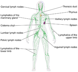
Группы лимфатических узлов.
Существует несколько групп лимфатических узлов. Располагаются эти группы таким образом, чтобы стать преградой на пути у инфекции и рака. Так, лимфоузлы располагаются в локтевом сгибе, подмышечной впадине, в коленном сгибе, а также паховой области. Лимфоузлы шеи обеспечивают защиту от инфекций и опухолей головы и органов, расположенных в области шеи. Огромное количество лимфатических узлов находится в брюшной и грудной полости. Лимфокапилляры пронизывают органы также как и поверхностные ткани. Лимфоузлы, располагающиеся по ходу кровеносных сосудов, выполняют те же самые функции.
На рисунке изображены следующие группы лимфатических узлов (сверху вниз):
- кольцо Вальдейера (Waldeyer ring) (глотка),
- шейные лимфатические узлы (Cervical),
- надключичные (supraclavicular),
- затылочные (occipital),
- передние ушные (preauricular),
- подключичные (Infraclavicular),
- подмышечные (Axillary),
- грудные (pectoral),
- внутригрудные, медиастинальные (Mediastinal),
- бронхопульмональные (Hilar),
- локтевые (Epitrochlear and brachial),
- селезёночные (Spleen),
- парааортальные (Paraaortic),
- брыжеечные (Mesenteric) (брыжейка)
- подвздошные (Iliac: общие, внутренние и внешние)
- паховые (Inguinal: глубокие и поверхностные),
- бедренные (femoral),
- подколенные (Popliteal).
Увеличение лимфатических узлов при инфекционных заболеваниях
Увеличение лимфатических узлов свидетельствует о неблагополучии в зоне, которую «обслуживает» узел. Чаще всего увеличение лимфоузла связано с инфекцией, реже оно является следствием опухолевого поражения.
- При гнойных процессах, как правило, возникает острый лимфаденит — воспаление лимфатического узла. Возникает воспалительный процесс вследствие попадания микробов из ран, расположенных в «зоне обслуживания» лимфоузла. Основным проявлением является увеличение лимфоузла, появление болезненности при его ощупывании. При возникновении гнойного процесса над лимфатическим узлом может краснеть кожа. Если в этот момент не вскрыть образовавшуюся полость, оболочка лимфоузла разрывается и гной проникает в окружающие ткани. Возникает тяжелое осложнение лимфаденита — флегмона.
- У детей увеличение лимфатических узлов при туберкулезе является одним из характерных проявлений инфекции. Чаще всего увеличиваются лимфоузлы грудной полости. Реже отмечается увеличение лимфоузлов шеи (в народе называют «золотухой»).
- Нередкой причиной увеличения лимфоузла у детей является болезнь кошачьей царапины. Возбудителем этой инфекции является микроб, называемый Бартонелла. Переносчиками бактерии являются кошки. Из царапины микробы распространяются по лимфатическим сосудам и попадают в лимфоузлы, которые увеличиваются и становятся болезненными. Незаживающая гнойная рана и увеличенный близлежащий лимфатический узел всегда должны наводить на мысль о болезни кошачьей царапины, как о причине такого состояния.
- При острых респираторных вирусных инфекциях (ОРВИ) у детей может отмечаться увеличение нескольких групп лимфоузлов. Является это следствием избыточного ответа иммунной системы на вторжение в организм вирусов. Как правило, лимфоузлы в таких случаях увеличиваются незначительно и при ощупывании болезненны.
- При венерических заболеваниях, в частности при сифилисе, увеличению лимфатического узла, как правило, в паховой области, предшествует возникновение язвы на половых органах — твердого шанкра. В отличие от других инфекционных заболеваний при сифилисе увеличенный лимфоузел может быть безболезненным.
- Длительно существующее увеличение нескольких групп лимфатических узлов может свидетельствовать о таких заболеваниях, как бруцеллез, листериоз, мононуклеоз, а также ВИЧ-инфекция.
Увеличение лимфоузлов при опухолевых заболеваниях
Опухолевое поражение лимфатических узлов может быть следствием как лимфопролиферативных заболеваний, когда первоначально опухоль исходит из лимфоузла, так и следствием метастатического поражения. К лимфопролиферативным заболеваниям относится, прежде всего, лимфогранулематоз и лимфосаркомы. Лимфоузлы при этих заболеваниях увеличиваются до 3-4 см, а иногда и больше, при этом становятся плотными. При ощупывании такие лимфатические узлы безболезненны. При первоначальном увеличении внутригрудных и внутрибрюшных лимфоузлов лимфопролиферативные заболевания могут быть распознаны не сразу.
Библиография
А. Г. Рахманова, В. К. Пригожкина, В. А. Неверов. Инфекционные болезни. Руководство для врачей общей практики. Москва-Санкт-Петербург, 1995.
Ссылки
- Лимфатический узел. Микрофотография.
- Эхограмма лимфатического узла
- Строение и функции лимфатического узла. Поражение лимфатических узлов и спленомегалия. // Справочник Харрисона по внутренним болезням
- Х. Э. Шайхова, А. М. Хакимов, В. А. Хорошаев Морфология регионарных лимфатических узлов при лимфотропной терапии острого среднего отита
- Лимфоузлы // Ленинградский областной онкологический диспансер
Wikimedia Foundation.
2010.
лимфати́ческий
Правильное ударение в этом слове падает на 3-й слог. На букву и
Посмотреть все слова на букву Л
Лимфатическая система состоит из небольших “узлов”, сложенных лимфатической тканью. Лимфоузлы — важная часть иммунитета. Если человек заболевает, то это можно обнаружить, проверив лимфоузлы, которые в таких ситуациях обычно опухают. Соответственно, если вы обнаружили у себя опухший лимфоузел, то есть смысл обратиться к врачу. Эта статья расскажет вам, как надо проверять лимфоузлы.
Метод 1
Проверка лимфоузлов
Знайте месторасположение лимфоузлов. Больше всего их в шее и в районе ключиц, в подмышках и в паху.
Лимфоузлы объединены в группы из нескольких узлов, размеры которых варьируются от размера горошины до размера боба.
Лимфоузлы, находящиеся в районе паха, так и называются — паховые.
Прижмите три пальца друг к другу, как показано на рисунке. Кончиками пальцев вы будете пальпировать себя — легонько нажимать на различные участки тела, где расположены лимфоузлы.
Надавите пальцами на предплечье. Запомните это ощущение — так ощущается здоровая, не отекшая и не опухшая область.
Теперь переместите руку в область подмышки и пропальпируйте там.Лимфоузлы находятся рядом с ребрами, в нижней части подмышки.
Пальпируйте аккуратно. Чувствуете что-то необычное? Вы должны чувствовать кости ребер, мышцы и, возможно, жир. Если же вы нащупали что-то припухшее и чувствительное, то это вполне может быть воспалившийся лимфоузел.
Второй рукой пропальпируйте вторую подмышку.
Размер отекших лимфоузлов часто такой же, как у горошин или фасоли.
Пропальпируйте лимфоузлы на шее и ключице. Кончиками пальцев обеих рук проведите круговыми движениями за ушами, потом спускайтесь к шее, затем пропальпируйте область под линией челюсти. Если вы нащупали что-то опухшее и болезненное — это может быть воспалившийся лимфоузел. К слову, воспаление лимфоузлов в этой области может также сопровождаться затрудненным глотанием и болью в горле.
Пропальпируйте область паха. Помните: мышцы, жир и кости — хорошо. Шишечки и опухоли — плохо.
Метод 2
Когда обращаться к врачу?
Следите за состоянием опухших лимфоузлов. Порой лимфоузлы воспаляются из-за аллергии, но это проходит за несколько дней. Если же прошло уже достаточно времени, а лимфоузлы вас по-прежнему беспокоят, нужно обратиться к врачу.
Проанализируйте состояние своего здоровья и подумайте, какие еще симптомы у вас наблюдаются. Воспалившийся лимфоузел означает, что организм борется с какой-то болезнью, возможно, даже с очень серьезной болезнью. Соответственно, если лимфоузлы воспалились на фоне одного из нижеследующих симптомов, то нужно немедленно обратиться к врачу! Итак, вот на что следует обратить внимание:
Необъяснимая потеря веса
Ночная потливость
Повышенная температура
Затрудненное дыхание, проблемы с проглатыванием
Узнайте свой диагноз. Врач, к которому вы придете с жалобами, проведет вас через все круги лабораторных исследований, выявит причину болезни и назначит программу лечения. Прямо сейчас можно сказать лишь то, что чаще всего лимфоузлы воспаляются из-за:
Инфекций (бактериальных и вирусных).
Проблем с иммунитетом.
Различных онкологических заболеваний.
Советы
Отекшие лимфоузлы — это не всегда симптом болезни. Если вы нехорошо себя чувствуете, если у вас отекли лимфоузлы, а симптомы что-то совсем не похожи на грипп или простуду — обратитесь к врачу.
Часто лимфоузлы за несколько дней возвращаются в норму сами по себе.
Предупреждения
Если в районе груди у вас нащупалась безболезненная опухоль — обратитесь к врачу.
Если воспаление лимфоузлов не проходит уже больше недели, либо если развились и другие симптомы (жар, озноб), то обратитесь к врачу.
Ещё больше полезной информации здесь
Источник
Лимфоузел
- Лимфоузел
-
1. Малая медицинская энциклопедия. — М.: Медицинская энциклопедия. 1991—96 гг. 2. Первая медицинская помощь. — М.: Большая Российская Энциклопедия. 1994 г. 3. Энциклопедический словарь медицинских терминов. — М.: Советская энциклопедия. — 1982—1984 гг.
Синонимы:
Смотреть что такое «Лимфоузел» в других словарях:
-
лимфоузел — сущ., кол во синонимов: 1 • узел (39) Словарь синонимов ASIS. В.Н. Тришин. 2013 … Словарь синонимов
-
Лимфоузел — Структура лимфатического узла. Лимфатический узел (лимфоузел) периферический орган лимфатической системы, выполняющий функцию биологического фильтра, через который протекает лимфа, поступающая от органов и частей тела. В теле человека выделяют… … Википедия
-
лимфоузел — (lymphonodus) см. Лимфатический узел … Большой медицинский словарь
-
Лимфаденопатия — МКБ 10 I88.88., L04.04., R59.159.1 МКБ 9 … Википедия
-
Лимфатический узел — Структура лимфатического узла. Лимфатический узел (лимфоузел) периферический орган лимфатической системы, выполняющий функцию биологического фильтра, через который протекает лимфа, поступающая от органов и частей … Википедия
-
Лимфатические узлы — Структура лимфатического узла. Лимфатический узел (лимфоузел) периферический орган лимфатической системы, выполняющий функцию биологического фильтра, через который протекает лимфа, поступающая от органов и частей тела. В теле человека выделяют… … Википедия
-
Лимфоузлы — Структура лимфатического узла. Лимфатический узел (лимфоузел) периферический орган лимфатической системы, выполняющий функцию биологического фильтра, через который протекает лимфа, поступающая от органов и частей тела. В теле человека выделяют… … Википедия
-
Меланома — МКБ 10 C … Википедия
-
лимфатический — ( ие) узел (узлы) (nodi lymphatici, PNA; lymphonodi, PNA, JNA; lymphoglandulae, BNA; син.: железа лимфатическая нрк, лимфоузел) общее название органов лимфатической системы, представляющих собой округлые мягкие образования, расположенные по ходу… … Большой медицинский словарь
-
Медиастиноскопия — (от новолатинского mediastinum средостение и …скопия (См. …скопия)) осмотр переднего средостения с целью биопсии (См. Биопсия). М. проводят в операционной под наркозом. Прибор для проведения М. медиастиноскоп полая трубка длиной 15 см с … Большая советская энциклопедия
-
Лимфати́ческий у́зел — ( ие) (узлы) (nodi lymphatici, PNA; lymphonodi. PNA, JNA; lymphoglandulae, BNA; сии.: железа лимфатическая нрк, лимфоузел) общее название органов лимфатической системы, представляющих собой округлые мягкие образования, расположенные по ходу… … Медицинская энциклопедия
| Lymph node | |
|---|---|

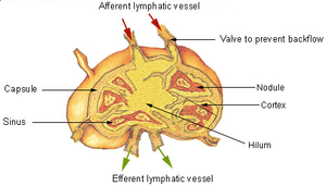
Diagram showing major parts of a lymph node. |
|
Lymph nodes form part of the lymphatic system, and are present in most parts of the body, and connected by small lymphatic vessels. |
|
| Details | |
| System | Lymphatic system, part of the immune system |
| Identifiers | |
| Latin | nodus lymphaticus (singular); nodi lymphatici (plural) |
| MeSH | D008198 |
| TA98 | A13.2.03.001 |
| TA2 | 5192 |
| FMA | 5034 |
| Anatomical terminology
[edit on Wikidata] |
A lymph node, or lymph gland,[1] is a kidney-shaped organ of the lymphatic system and the adaptive immune system. A large number of lymph nodes are linked throughout the body by the lymphatic vessels. They are major sites of lymphocytes that include B and T cells. Lymph nodes are important for the proper functioning of the immune system, acting as filters for foreign particles including cancer cells, but have no detoxification function.
In the lymphatic system a lymph node is a secondary lymphoid organ. A lymph node is enclosed in a fibrous capsule and is made up of an outer cortex and an inner medulla.
Lymph nodes become inflamed or enlarged in various diseases, which may range from trivial throat infections to life-threatening cancers. The condition of lymph nodes is very important in cancer staging, which decides the treatment to be used and determines the prognosis. Lymphadenopathy refers to glands that are enlarged or swollen. When inflamed or enlarged, lymph nodes can be firm or tender.
Structure[edit]
Cross-section of a lymph node with sections labelled.1) Capsule; 2) Subcapsular sinus; 3) Germinal centre; 4) Lymphoid nodule; 5) Trabeculae
Lymph nodes are kidney or oval shaped and range in size from 2 mm to 25 mm on their long axis, with an average of 15 mm.[2]
Each lymph node is surrounded by a fibrous capsule, which extends inside a lymph node to form trabeculae.[3] The substance of a lymph node is divided into the outer cortex and the inner medulla.[3] These are rich with cells.[4] The hilum is an indent on the concave surface of the lymph node where lymphatic vessels leave and blood vessels enter and leave.[4]
Lymph enters the convex side of a lymph node through multiple afferent lymphatic vessels and from there flows into a series of sinuses.[3] After entering the lymph node from afferent lymphatic vessels, lymph flows into a space underneath the capsule called the subcapsular sinus, then into cortical sinuses.[3] After passing through the cortex, lymph then collects in medullary sinuses.[3] All of these sinuses drain into the efferent lymph vessels to exit the node at the hilum on the concave side.[3]
Location[edit]
Lymph nodes are present throughout the body, are more concentrated near and within the trunk, and are divided into groups.[4] There are about 450 lymph nodes in the adult.[4] Some lymph nodes can be felt when enlarged (and occasionally when not), such as the axillary lymph nodes under the arm, the cervical lymph nodes of the head and neck and the inguinal lymph nodes near the groin crease. Most lymph nodes lie within the trunk adjacent to other major structures in the body — such as the paraaortic lymph nodes and the tracheobronchial lymph nodes. The lymphatic drainage patterns are different from person to person and even asymmetrical on each side of the same body.[5][6]
There are no lymph nodes in the central nervous system, which is separated from the body by the blood–brain barrier. Lymph from the meningeal lymphatic vessels in the CNS drains to the deep cervical lymph nodes.[7]
Size[edit]
| Generally | 10 mm[8][9] |
| Inguinal | 10[10] – 20 mm[11] |
| Pelvis | 10 mm for ovoid lymph nodes, 8 mm for rounded[10] |
| Neck | |
|---|---|
| Generally (non-retropharyngeal) | 10 mm[10][12] |
| Jugulodigastric lymph nodes | 11mm[10] or 15 mm[12] |
| Retropharyngeal | 8 mm[12]
|
| Mediastinum | |
| Mediastinum, generally | 10 mm[10] |
| Superior mediastinum and high paratracheal | 7mm[13] |
| Low paratracheal and subcarinal | 11 mm[13] |
| Upper abdominal | |
| Retrocrural space | 6 mm[14] |
| Paracardiac | 8 mm[14] |
| Gastrohepatic ligament | 8 mm[14] |
| Upper paraaortic region | 9 mm[14] |
| Portacaval space | 10 mm[14] |
| Porta hepatis | 7 mm[14] |
| Lower paraaortic region | 11 mm[14] |
Subdivisions[edit]
Histology of a normal lymphoid follicle, showing dark, light, mantle and marginal zones.
A lymph node is divided into compartments called nodules (or lobules), each consisting of a region of cortex with combined follicle B cells, a paracortex of T cells, and a part of the nodule in the medulla.[15] The substance of a lymph node is divided into the outer cortex and the inner medulla.[3] The cortex of a lymph node is the outer portion of the node, underneath the capsule and the subcapsular sinus.[15] It has an outer part and a deeper part known as the paracortex.[15] The outer cortex consists of groups of mainly inactivated B cells called follicles.[4] When activated, these may develop into what is called a germinal centre.[4] The deeper paracortex mainly consists of the T cells.[4] Here the T-cells mainly interact with dendritic cells, and the reticular network is dense.[16]
The medulla contains large blood vessels, sinuses and medullary cords that contain antibody-secreting plasma cells. There are fewer cells in the medulla.[4]
The medullary cords are cords of lymphatic tissue, and include plasma cells, macrophages, and B cells.
Cells[edit]
In the lymphatic system a lymph node is a secondary lymphoid organ.[4] Lymph nodes contain lymphocytes, a type of white blood cell, and are primarily made up of B cells and T cells.[4] B cells are mainly found in the outer cortex where they are clustered together as follicular B cells in lymphoid follicles, and T cells and dendritic cells are mainly found in the paracortex.[17]
There are fewer cells in the medulla than the cortex.[4] The medulla contains plasma cells, as well as macrophages which are present within the medullary sinuses.[17]
As part of the reticular network, there are follicular dendritic cells in the B cell follicle and fibroblastic reticular cells in the T cell cortex. The reticular network provides structural support and a surface for adhesion of the dendritic cells, macrophages and lymphocytes. It also allows exchange of material with blood through the high endothelial venules and provides the growth and regulatory factors necessary for activation and maturation of immune cells.[18]
Lymph flow[edit]
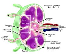
Labeled diagram of human lymph node showing the flow of lymph.
Afferent and efferent vessels
Lymph enters the convex side of a lymph node through multiple afferent lymphatic vessels, which form a network of lymphatic vessels (Latin: plexus) and from here flows into a space (Latin: sinus) underneath the capsule called the subcapsular sinus.[4][3] From here, lymph flows into sinuses within the cortex.[3] After passing through the cortex, lymph then collects in medullary sinuses.[3] All of these sinuses drain into the efferent lymphatic vessels to exit the node at the hilum on the concave side.[3]
These are channels within the node lined by endothelial cells along with fibroblastic reticular cells, allowing for the smooth flow of lymph. The endothelium of the subcapsular sinus is continuous with that of the afferent lymph vessel and also with that of the similar sinuses flanking the trabeculae and within the cortex. These vessels are smaller and don’t allow the passage of macrophages so that they remain contained to function within a lymph node. In the course of the lymph, lymphocytes may be activated as part of the adaptive immune response.
There is usually only one efferent vessel though sometimes there may be two.[19] Medullary sinuses contain histiocytes (immobile macrophages) and reticular cells.
A lymph node contains lymphoid tissue, i.e., a meshwork or fibers called reticulum with white blood cells enmeshed in it. The regions where there are few cells within the meshwork are known as lymph sinus. It is lined by reticular cells, fibroblasts and fixed macrophages.[20]
Capsule[edit]
Lymph node tissue showing trabeculae
Thin reticular fibers (reticulin) of reticular connective tissue form a supporting meshwork inside the node.[4]
The lymph node capsule is composed of dense irregular connective tissue with some plain collagenous fibers, and a number of membranous processes or trabeculae extend from its internal surface. The trabeculae pass inward, radiating toward the center of the node, for about one-third or one-fourth of the space between the circumference and the center of the node. In some animals they are sufficiently well-marked to divide the peripheral or cortical portion of the node into a number of compartments (nodules), but in humans this arrangement is not obvious. The larger trabeculae springing from the capsule break up into finer bands, and these interlace to form a mesh-work in the central or medullary portion of the node. These trabecular spaces formed by the interlacing trabeculae contain the proper lymph node substance or lymphoid tissue. The node pulp does not, however, completely fill the spaces, but leaves between its outer margin and the enclosing trabeculae a channel or space of uniform width throughout. This is termed the subcapsular sinus (lymph path or lymph sinus). Running across it are a number of finer trabeculae of reticular fibers, mostly covered by ramifying cells.
Function[edit]
In the lymphatic system a lymph node is a secondary lymphoid organ.[4]
The primary function of lymph nodes is the filtering of lymph to identify and fight infection. In order to do this, lymph nodes contain lymphocytes, a type of white blood cell, which includes B cells and T cells. These circulate through the bloodstream and enter and reside in lymph nodes.[21] B cells produce antibodies. Each antibody has a single predetermined target, an antigen, that it can bind to. These circulate throughout the bloodstream and if they find this target, the antibodies bind to it and stimulate an immune response. Each B cell produces different antibodies, and this process is driven in lymph nodes. B cells enter the bloodstream as «naive» cells produced in bone marrow. After entering a lymph node, they then enter a lymphoid follicle, where they multiply and divide, each producing a different antibody. If a cell is stimulated, it will go on to produce more antibodies (a plasma cell) or act as a memory cell to help the body fight future infection.[22] If a cell is not stimulated, it will undergo apoptosis and die.[22]
Antigens are molecules found on bacterial cell walls, chemical substances secreted from bacteria, or sometimes even molecules present in body tissue itself. These are taken up by cells throughout the body called antigen-presenting cells, such as dendritic cells.[23] These antigen presenting cells enter the lymph system and then lymph nodes. They present the antigen to T cells and, if there is a T cell with the appropriate T cell receptor, it will be activated.[22]
B cells acquire antigen directly from the afferent lymph. If a B cell binds its cognate antigen it will be activated. Some B cells will immediately develop into antibody secreting plasma cells, and secrete IgM. Other B cells will internalize the antigen and present it to follicular helper T cells on the B and T cell zone interface. If a cognate FTh cell is found it will upregulate CD40L and promote somatic hypermutation and isotype class switching of the B cell, increasing its antigen binding affinity and changing its effector function. Proliferation of cells within a lymph node will make the node expand.
Lymph is present throughout the body, and circulates through lymphatic vessels. These drain into and from lymph nodes – afferent vessels drain into nodes, and efferent vessels from nodes. When lymph fluid enters a node, it drains into the node just beneath the capsule in a space called the subcapsular sinus. The subcapsular sinus drains into trabecular sinuses and finally into medullary sinuses. The sinus space is criss-crossed by the pseudopods of macrophages, which act to trap foreign particles and filter the lymph. The medullary sinuses converge at the hilum and lymph then leaves the lymph node via the efferent lymphatic vessel towards either a more central lymph node or ultimately for drainage into a central venous subclavian blood vessel.
- The B cells migrate to the nodular cortex and medulla.
- The T cells migrate to the deep cortex. This is a region of a lymph node called the paracortex that immediately surrounds the medulla. Because both naive T cells and dendritic cells express CCR7, they are drawn into the paracortex by the same chemotactic factors, increasing the chance of T cell activation. Both B and T lymphocytes enter lymph nodes from circulating blood through specialized high endothelial venules found in the paracortex.
Clinical significance[edit]
Swelling[edit]
A still image from a 3D medical animation showing enlarged lymph nodes.
Lymph node enlargement or swelling is known as lymphadenopathy.[24] Swelling may be due to many causes, including infections, tumors, autoimmune disease, drug reactions, diseases such as amyloidosis and sarcoidosis, or because of lymphoma or leukemia.[25][24] Depending on the cause, swelling may be painful, particularly if the expansion is rapid and due to an infection or inflammation.[24] Lymph node enlargement may be localised to an area, which might suggest a local source of infection or a tumour in that area that has spread to the lymph node.[24] It may also be generalised, which might suggest infection, connective tissue or autoimmune disease, or a malignancy of blood cells such as a lymphoma or leukaemia.[24] Rarely, depending on location, lymph node enlargement may cause problems such as difficulty breathing, or compression of a blood vessel (for example, superior vena cava obstruction[26]).
Enlarged lymph nodes might be felt as part of a medical examination, or found on medical imaging.[27] Features of the medical history may point to the cause, such as the speed of onset of swelling, pain, and other constitutional symptoms such as fevers or weight loss.[28] For example, a tumour of the breast may result in swelling of the lymph nodes under the arms[24] and weight loss and night sweats may suggest a malignancy such as lymphoma.[24]
In addition to a medical exam by a medical practitioner, medical tests may include blood tests and scans may be needed to further examine the cause.[24] A biopsy of a lymph node may also be needed.[24]
Cancer[edit]
Lymph nodes can be affected by both primary cancers of lymph tissue, and secondary cancers affecting other parts of the body. Primary cancers of lymph tissue are called lymphomas and include Hodgkin lymphoma and non-Hodgkin lymphoma.[29] Cancer of lymph nodes can cause a wide range of symptoms from painless long-term slowly growing swelling to sudden, rapid enlargement over days or weeks, with symptoms depending on the grade of the tumour.[29] Most lymphomas are tumours of B-cells.[29] Lymphoma is managed by haematologists and oncologists.
Local cancer in many parts of the body can cause lymph nodes to enlarge because of tumorous cells that have metastasised into the node.[30] Lymph node involvement is often a key part in the diagnosis and treatment of cancer, acting as «sentinels» of local disease, incorporated into TNM staging and other cancer staging systems. As part of the investigations or workup for cancer, lymph nodes may be imaged or even surgically removed. If removed, the lymph node will be stained and examined under a microscope by a pathologist to determine if there is evidence of cells that appear cancerous (i.e. have metastasized into the node). The staging of the cancer, and therefore the treatment approach and prognosis, is predicated on the presence of node metastases.
Lymphedema[edit]
Lymphedema is the condition of swelling (edema) of tissue relating to insufficient clearance by the lymphatic system.[31] It can be congenital as a result usually of undeveloped or absent lymph nodes, and is known as primary lymphedema. Lymphedema most commonly arises in the arms or legs, but can also occur in the chest wall, genitals, neck, and abdomen.[32] Secondary lymphedema usually results from the removal of lymph nodes during breast cancer surgery or from other damaging treatments such as radiation. It can also be caused by some parasitic infections. Affected tissues are at a great risk of infection.[citation needed] Management of lymphedema may include advice to lose weight, exercise, keep the affected limb moist, and compress the affected area.[31] Sometimes surgical management is also considered.[31]
Similar lymphoid organs[edit]
The spleen and the tonsils are the larger secondary lymphoid organs that serve somewhat similar functions to lymph nodes, though the spleen filters blood cells rather than lymph. The tonsils are sometimes erroneously referred to as lymph nodes. Although the tonsils and lymph nodes do share certain characteristics, there are also many important differences between them, such as their location, structure and size.[33] Furthermore, the tonsils filter tissue fluid whereas lymph nodes filter lymph.[33]
The appendix contains lymphoid tissue and is therefore believed to play a role not only in the digestive system, but also in the immune system.[34]
See also[edit]
- Peyer’s patch
- Lymph sacs
References[edit]
- ^ «Swollen glands NHS inform». www.nhsinform.scot. Retrieved 4 April 2020.
- ^ Ioachim, Harry L. (1994). Lymph Node Pathology (Second ed.). J. B. Lippincott Company. p. 3. ISBN 9780397508075. Retrieved 17 October 2022.
- ^ a b c d e f g h i j k Young B, O’Dowd G, Woodford P (2013). Wheater’s functional histology: a text and colour atlas (6th ed.). Philadelphia: Elsevier. pp. 209–210. ISBN 9780702047473.
- ^ a b c d e f g h i j k l m n Standring, Susan, ed. (2016). «Lymphoid tissues». Gray’s anatomy : the anatomical basis of clinical practice (41st ed.). Philadelphia. pp. 73–4. ISBN 9780702052309. OCLC 920806541.
- ^ Themes, U. F. O. (6 January 2018). «Lymphatic Anatomy and Clinical Implications». Ento Key. Retrieved 21 September 2020.
- ^ Pan, Wei-Ren; Wang, De-Guang (2013). «Historical review of lymphatic studies in the head and neck» (PDF). Journal of Lymphoedema. WoundsGroup. 8.
- ^ Dupont G, Schmidt C, Yilmaz E, Oskouian RJ, Macchi V, de Caro R, Tubbs RS (January 2019). «Our current understanding of the lymphatics of the brain and spinal cord». Clinical Anatomy. 32 (1): 117–121. doi:10.1002/ca.23308. PMID 30362622.
- ^ Ganeshalingam, Skandadas; Koh, Dow-Mu (2009). «Nodal staging». Cancer Imaging. 9 (1): 104–111. doi:10.1102/1470-7330.2009.0017. ISSN 1470-7330. PMC 2821588. PMID 20080453.
- ^ Schmidt Júnior, Aurelino Fernandes; Rodrigues, Olavo Ribeiro; Matheus, Roberto Storte; Kim, Jorge Du Ub; Jatene, Fábio Biscegli (2007). «Distribuição, tamanho e número dos linfonodos mediastinais: definições por meio de estudo anatômico». Jornal Brasileiro de Pneumologia. 33 (2): 134–140. doi:10.1590/S1806-37132007000200006. ISSN 1806-3713. PMID 17724531.
- ^ a b c d e f Torabi M, Aquino SL, Harisinghani MG (September 2004). «Current concepts in lymph node imaging». Journal of Nuclear Medicine. 45 (9): 1509–18. PMID 15347718.
- ^ «Assessment of lymphadenopathy». BMJ Best Practice. Retrieved 4 March 2017. Last updated: Last updated: Feb 16, 2017
- ^ a b c Page 432 in: Luca Saba (2016). Image Principles, Neck, and the Brain. CRC Press. ISBN 9781482216202.
- ^ a b Sharma, Amita; Fidias, Panos; Hayman, L. Anne; Loomis, Susanne L.; Taber, Katherine H.; Aquino, Suzanne L. (2004). «Patterns of Lymphadenopathy in Thoracic Malignancies». RadioGraphics. 24 (2): 419–434. doi:10.1148/rg.242035075. ISSN 0271-5333. PMID 15026591. S2CID 7434544.
- ^ a b c d e f g Dorfman, R E; Alpern, M B; Gross, B H; Sandler, M A (1991). «Upper abdominal lymph nodes: criteria for normal size determined with CT». Radiology. 180 (2): 319–322. doi:10.1148/radiology.180.2.2068292. ISSN 0033-8419. PMID 2068292.
- ^ a b c Willard-Mack CL (25 June 2016). «Normal structure, function, and histology of lymph nodes». Toxicologic Pathology. 34 (5): 409–24. doi:10.1080/01926230600867727. PMID 17067937.
- ^ Katakai T, Hara T, Lee JH, Gonda H, Sugai M, Shimizu A (August 2004). «A novel reticular stromal structure in lymph node cortex: an immuno-platform for interactions among dendritic cells, T cells and B cells». International Immunology. 16 (8): 1133–42. doi:10.1093/intimm/dxh113. PMID 15237106.
- ^ a b Davidson’s 2018, p. 67.
- ^ Kaldjian EP, Gretz JE, Anderson AO, Shi Y, Shaw S (October 2001). «Spatial and molecular organization of lymph node T cell cortex: a labyrinthine cavity bounded by an epithelium-like monolayer of fibroblastic reticular cells anchored to basement membrane-like extracellular matrix». International Immunology. 13 (10): 1243–53. doi:10.1093/intimm/13.10.1243. PMID 11581169.
- ^ Henrikson RC, Mazurkiewicz JE (1 January 1997). Histology. Lippincott Williams & Wilkins. ISBN 9780683062250.
- ^ Warwick R, Williams PL (1973) [1858]. «Angiology (Chapter 6)». Gray’s anatomy. illustrated by Richard E. M. Moo re (Thirty-fifth ed.). London: Longman. pp. 588–785.
- ^ Hoffbrand’s 2016, p. 103,110.
- ^ a b c Hoffbrand’s 2016, p. 111.
- ^ Hoffbrand’s 2016, p. 109.
- ^ a b c d e f g h i Davidson’s 2018, p. 927.
- ^ Hoffbrand’s 2016, p. 114.
- ^ Davidson’s 2018, p. 1326.
- ^ Gaddey, Heidi L.; Riegel, Angela M. (1 December 2016). «Unexplained Lymphadenopathy: Evaluation and Differential Diagnosis». American Family Physician. 94 (11): 896–903. ISSN 1532-0650. PMID 27929264.
- ^ Davidson’s 2018, p. 913.
- ^ a b c Davidson’s 2018, p. 961.
- ^ Davidson’s 2018, p. 1324.
- ^ a b c Maclellan RA, Greene AK (August 2014). «Lymphedema». Seminars in Pediatric Surgery. 23 (4): 191–7. doi:10.1053/j.sempedsurg.2014.07.004. PMID 25241097.
- ^ «Lymphedema». MayoClinic.org. 18 September 2019. Retrieved 17 November 2022.
- ^ a b Lakna (31 January 2019). «What is the Difference Between Tonsils and Lymph Nodes». Pediaa.Com. Retrieved 14 December 2019.
- ^ Kooij IA, Sahami S, Meijer SL, Buskens CJ, Te Velde AA (October 2016). «The immunology of the vermiform appendix: a review of the literature». Clinical and Experimental Immunology. 186 (1): 1–9. doi:10.1111/cei.12821. PMC 5011360. PMID 27271818.
Bibliography[edit]
- Ralston SH, Penman ID, Strachan MW, Hobson RP (2018). Davidson’s principles and practice of medicine (23rd ed.). Elsevier. ISBN 978-0-7020-7028-0.
- Hoffbrand V, Moss PA (2016). Hoffbrand’s essential haematology (7th ed.). West Sussex: Wiley Blackwell. ISBN 978-1-1184-0867-4.
External links[edit]
- Histology image: 07101loa – Histology Learning System at Boston University
- Lymph Nodes
- Lymph Nodes Drainage
- An overview of Normal Lymph Nodes and Swollen lymph nodes and their evaluation
| Lymph node | |
|---|---|

Diagram showing major parts of a lymph node. |
|

Lymph nodes form part of the lymphatic system, and are present in most parts of the body, and connected by small lymphatic vessels. |
|
| Details | |
| System | Lymphatic system, part of the immune system |
| Identifiers | |
| Latin | nodus lymphaticus (singular); nodi lymphatici (plural) |
| MeSH | D008198 |
| TA98 | A13.2.03.001 |
| TA2 | 5192 |
| FMA | 5034 |
| Anatomical terminology
[edit on Wikidata] |
A lymph node, or lymph gland,[1] is a kidney-shaped organ of the lymphatic system and the adaptive immune system. A large number of lymph nodes are linked throughout the body by the lymphatic vessels. They are major sites of lymphocytes that include B and T cells. Lymph nodes are important for the proper functioning of the immune system, acting as filters for foreign particles including cancer cells, but have no detoxification function.
In the lymphatic system a lymph node is a secondary lymphoid organ. A lymph node is enclosed in a fibrous capsule and is made up of an outer cortex and an inner medulla.
Lymph nodes become inflamed or enlarged in various diseases, which may range from trivial throat infections to life-threatening cancers. The condition of lymph nodes is very important in cancer staging, which decides the treatment to be used and determines the prognosis. Lymphadenopathy refers to glands that are enlarged or swollen. When inflamed or enlarged, lymph nodes can be firm or tender.
Structure[edit]
Cross-section of a lymph node with sections labelled.1) Capsule; 2) Subcapsular sinus; 3) Germinal centre; 4) Lymphoid nodule; 5) Trabeculae
Lymph nodes are kidney or oval shaped and range in size from 2 mm to 25 mm on their long axis, with an average of 15 mm.[2]
Each lymph node is surrounded by a fibrous capsule, which extends inside a lymph node to form trabeculae.[3] The substance of a lymph node is divided into the outer cortex and the inner medulla.[3] These are rich with cells.[4] The hilum is an indent on the concave surface of the lymph node where lymphatic vessels leave and blood vessels enter and leave.[4]
Lymph enters the convex side of a lymph node through multiple afferent lymphatic vessels and from there flows into a series of sinuses.[3] After entering the lymph node from afferent lymphatic vessels, lymph flows into a space underneath the capsule called the subcapsular sinus, then into cortical sinuses.[3] After passing through the cortex, lymph then collects in medullary sinuses.[3] All of these sinuses drain into the efferent lymph vessels to exit the node at the hilum on the concave side.[3]
Location[edit]
Lymph nodes are present throughout the body, are more concentrated near and within the trunk, and are divided into groups.[4] There are about 450 lymph nodes in the adult.[4] Some lymph nodes can be felt when enlarged (and occasionally when not), such as the axillary lymph nodes under the arm, the cervical lymph nodes of the head and neck and the inguinal lymph nodes near the groin crease. Most lymph nodes lie within the trunk adjacent to other major structures in the body — such as the paraaortic lymph nodes and the tracheobronchial lymph nodes. The lymphatic drainage patterns are different from person to person and even asymmetrical on each side of the same body.[5][6]
There are no lymph nodes in the central nervous system, which is separated from the body by the blood–brain barrier. Lymph from the meningeal lymphatic vessels in the CNS drains to the deep cervical lymph nodes.[7]
Size[edit]
| Generally | 10 mm[8][9] |
| Inguinal | 10[10] – 20 mm[11] |
| Pelvis | 10 mm for ovoid lymph nodes, 8 mm for rounded[10] |
| Neck | |
|---|---|
| Generally (non-retropharyngeal) | 10 mm[10][12] |
| Jugulodigastric lymph nodes | 11mm[10] or 15 mm[12] |
| Retropharyngeal | 8 mm[12]
|
| Mediastinum | |
| Mediastinum, generally | 10 mm[10] |
| Superior mediastinum and high paratracheal | 7mm[13] |
| Low paratracheal and subcarinal | 11 mm[13] |
| Upper abdominal | |
| Retrocrural space | 6 mm[14] |
| Paracardiac | 8 mm[14] |
| Gastrohepatic ligament | 8 mm[14] |
| Upper paraaortic region | 9 mm[14] |
| Portacaval space | 10 mm[14] |
| Porta hepatis | 7 mm[14] |
| Lower paraaortic region | 11 mm[14] |
Subdivisions[edit]
Histology of a normal lymphoid follicle, showing dark, light, mantle and marginal zones.
A lymph node is divided into compartments called nodules (or lobules), each consisting of a region of cortex with combined follicle B cells, a paracortex of T cells, and a part of the nodule in the medulla.[15] The substance of a lymph node is divided into the outer cortex and the inner medulla.[3] The cortex of a lymph node is the outer portion of the node, underneath the capsule and the subcapsular sinus.[15] It has an outer part and a deeper part known as the paracortex.[15] The outer cortex consists of groups of mainly inactivated B cells called follicles.[4] When activated, these may develop into what is called a germinal centre.[4] The deeper paracortex mainly consists of the T cells.[4] Here the T-cells mainly interact with dendritic cells, and the reticular network is dense.[16]
The medulla contains large blood vessels, sinuses and medullary cords that contain antibody-secreting plasma cells. There are fewer cells in the medulla.[4]
The medullary cords are cords of lymphatic tissue, and include plasma cells, macrophages, and B cells.
Cells[edit]
In the lymphatic system a lymph node is a secondary lymphoid organ.[4] Lymph nodes contain lymphocytes, a type of white blood cell, and are primarily made up of B cells and T cells.[4] B cells are mainly found in the outer cortex where they are clustered together as follicular B cells in lymphoid follicles, and T cells and dendritic cells are mainly found in the paracortex.[17]
There are fewer cells in the medulla than the cortex.[4] The medulla contains plasma cells, as well as macrophages which are present within the medullary sinuses.[17]
As part of the reticular network, there are follicular dendritic cells in the B cell follicle and fibroblastic reticular cells in the T cell cortex. The reticular network provides structural support and a surface for adhesion of the dendritic cells, macrophages and lymphocytes. It also allows exchange of material with blood through the high endothelial venules and provides the growth and regulatory factors necessary for activation and maturation of immune cells.[18]
Lymph flow[edit]
Labeled diagram of human lymph node showing the flow of lymph.
Afferent and efferent vessels
Lymph enters the convex side of a lymph node through multiple afferent lymphatic vessels, which form a network of lymphatic vessels (Latin: plexus) and from here flows into a space (Latin: sinus) underneath the capsule called the subcapsular sinus.[4][3] From here, lymph flows into sinuses within the cortex.[3] After passing through the cortex, lymph then collects in medullary sinuses.[3] All of these sinuses drain into the efferent lymphatic vessels to exit the node at the hilum on the concave side.[3]
These are channels within the node lined by endothelial cells along with fibroblastic reticular cells, allowing for the smooth flow of lymph. The endothelium of the subcapsular sinus is continuous with that of the afferent lymph vessel and also with that of the similar sinuses flanking the trabeculae and within the cortex. These vessels are smaller and don’t allow the passage of macrophages so that they remain contained to function within a lymph node. In the course of the lymph, lymphocytes may be activated as part of the adaptive immune response.
There is usually only one efferent vessel though sometimes there may be two.[19] Medullary sinuses contain histiocytes (immobile macrophages) and reticular cells.
A lymph node contains lymphoid tissue, i.e., a meshwork or fibers called reticulum with white blood cells enmeshed in it. The regions where there are few cells within the meshwork are known as lymph sinus. It is lined by reticular cells, fibroblasts and fixed macrophages.[20]
Capsule[edit]
Lymph node tissue showing trabeculae
Thin reticular fibers (reticulin) of reticular connective tissue form a supporting meshwork inside the node.[4]
The lymph node capsule is composed of dense irregular connective tissue with some plain collagenous fibers, and a number of membranous processes or trabeculae extend from its internal surface. The trabeculae pass inward, radiating toward the center of the node, for about one-third or one-fourth of the space between the circumference and the center of the node. In some animals they are sufficiently well-marked to divide the peripheral or cortical portion of the node into a number of compartments (nodules), but in humans this arrangement is not obvious. The larger trabeculae springing from the capsule break up into finer bands, and these interlace to form a mesh-work in the central or medullary portion of the node. These trabecular spaces formed by the interlacing trabeculae contain the proper lymph node substance or lymphoid tissue. The node pulp does not, however, completely fill the spaces, but leaves between its outer margin and the enclosing trabeculae a channel or space of uniform width throughout. This is termed the subcapsular sinus (lymph path or lymph sinus). Running across it are a number of finer trabeculae of reticular fibers, mostly covered by ramifying cells.
Function[edit]
In the lymphatic system a lymph node is a secondary lymphoid organ.[4]
The primary function of lymph nodes is the filtering of lymph to identify and fight infection. In order to do this, lymph nodes contain lymphocytes, a type of white blood cell, which includes B cells and T cells. These circulate through the bloodstream and enter and reside in lymph nodes.[21] B cells produce antibodies. Each antibody has a single predetermined target, an antigen, that it can bind to. These circulate throughout the bloodstream and if they find this target, the antibodies bind to it and stimulate an immune response. Each B cell produces different antibodies, and this process is driven in lymph nodes. B cells enter the bloodstream as «naive» cells produced in bone marrow. After entering a lymph node, they then enter a lymphoid follicle, where they multiply and divide, each producing a different antibody. If a cell is stimulated, it will go on to produce more antibodies (a plasma cell) or act as a memory cell to help the body fight future infection.[22] If a cell is not stimulated, it will undergo apoptosis and die.[22]
Antigens are molecules found on bacterial cell walls, chemical substances secreted from bacteria, or sometimes even molecules present in body tissue itself. These are taken up by cells throughout the body called antigen-presenting cells, such as dendritic cells.[23] These antigen presenting cells enter the lymph system and then lymph nodes. They present the antigen to T cells and, if there is a T cell with the appropriate T cell receptor, it will be activated.[22]
B cells acquire antigen directly from the afferent lymph. If a B cell binds its cognate antigen it will be activated. Some B cells will immediately develop into antibody secreting plasma cells, and secrete IgM. Other B cells will internalize the antigen and present it to follicular helper T cells on the B and T cell zone interface. If a cognate FTh cell is found it will upregulate CD40L and promote somatic hypermutation and isotype class switching of the B cell, increasing its antigen binding affinity and changing its effector function. Proliferation of cells within a lymph node will make the node expand.
Lymph is present throughout the body, and circulates through lymphatic vessels. These drain into and from lymph nodes – afferent vessels drain into nodes, and efferent vessels from nodes. When lymph fluid enters a node, it drains into the node just beneath the capsule in a space called the subcapsular sinus. The subcapsular sinus drains into trabecular sinuses and finally into medullary sinuses. The sinus space is criss-crossed by the pseudopods of macrophages, which act to trap foreign particles and filter the lymph. The medullary sinuses converge at the hilum and lymph then leaves the lymph node via the efferent lymphatic vessel towards either a more central lymph node or ultimately for drainage into a central venous subclavian blood vessel.
- The B cells migrate to the nodular cortex and medulla.
- The T cells migrate to the deep cortex. This is a region of a lymph node called the paracortex that immediately surrounds the medulla. Because both naive T cells and dendritic cells express CCR7, they are drawn into the paracortex by the same chemotactic factors, increasing the chance of T cell activation. Both B and T lymphocytes enter lymph nodes from circulating blood through specialized high endothelial venules found in the paracortex.
Clinical significance[edit]
Swelling[edit]
A still image from a 3D medical animation showing enlarged lymph nodes.
Lymph node enlargement or swelling is known as lymphadenopathy.[24] Swelling may be due to many causes, including infections, tumors, autoimmune disease, drug reactions, diseases such as amyloidosis and sarcoidosis, or because of lymphoma or leukemia.[25][24] Depending on the cause, swelling may be painful, particularly if the expansion is rapid and due to an infection or inflammation.[24] Lymph node enlargement may be localised to an area, which might suggest a local source of infection or a tumour in that area that has spread to the lymph node.[24] It may also be generalised, which might suggest infection, connective tissue or autoimmune disease, or a malignancy of blood cells such as a lymphoma or leukaemia.[24] Rarely, depending on location, lymph node enlargement may cause problems such as difficulty breathing, or compression of a blood vessel (for example, superior vena cava obstruction[26]).
Enlarged lymph nodes might be felt as part of a medical examination, or found on medical imaging.[27] Features of the medical history may point to the cause, such as the speed of onset of swelling, pain, and other constitutional symptoms such as fevers or weight loss.[28] For example, a tumour of the breast may result in swelling of the lymph nodes under the arms[24] and weight loss and night sweats may suggest a malignancy such as lymphoma.[24]
In addition to a medical exam by a medical practitioner, medical tests may include blood tests and scans may be needed to further examine the cause.[24] A biopsy of a lymph node may also be needed.[24]
Cancer[edit]
Lymph nodes can be affected by both primary cancers of lymph tissue, and secondary cancers affecting other parts of the body. Primary cancers of lymph tissue are called lymphomas and include Hodgkin lymphoma and non-Hodgkin lymphoma.[29] Cancer of lymph nodes can cause a wide range of symptoms from painless long-term slowly growing swelling to sudden, rapid enlargement over days or weeks, with symptoms depending on the grade of the tumour.[29] Most lymphomas are tumours of B-cells.[29] Lymphoma is managed by haematologists and oncologists.
Local cancer in many parts of the body can cause lymph nodes to enlarge because of tumorous cells that have metastasised into the node.[30] Lymph node involvement is often a key part in the diagnosis and treatment of cancer, acting as «sentinels» of local disease, incorporated into TNM staging and other cancer staging systems. As part of the investigations or workup for cancer, lymph nodes may be imaged or even surgically removed. If removed, the lymph node will be stained and examined under a microscope by a pathologist to determine if there is evidence of cells that appear cancerous (i.e. have metastasized into the node). The staging of the cancer, and therefore the treatment approach and prognosis, is predicated on the presence of node metastases.
Lymphedema[edit]
Lymphedema is the condition of swelling (edema) of tissue relating to insufficient clearance by the lymphatic system.[31] It can be congenital as a result usually of undeveloped or absent lymph nodes, and is known as primary lymphedema. Lymphedema most commonly arises in the arms or legs, but can also occur in the chest wall, genitals, neck, and abdomen.[32] Secondary lymphedema usually results from the removal of lymph nodes during breast cancer surgery or from other damaging treatments such as radiation. It can also be caused by some parasitic infections. Affected tissues are at a great risk of infection.[citation needed] Management of lymphedema may include advice to lose weight, exercise, keep the affected limb moist, and compress the affected area.[31] Sometimes surgical management is also considered.[31]
Similar lymphoid organs[edit]
The spleen and the tonsils are the larger secondary lymphoid organs that serve somewhat similar functions to lymph nodes, though the spleen filters blood cells rather than lymph. The tonsils are sometimes erroneously referred to as lymph nodes. Although the tonsils and lymph nodes do share certain characteristics, there are also many important differences between them, such as their location, structure and size.[33] Furthermore, the tonsils filter tissue fluid whereas lymph nodes filter lymph.[33]
The appendix contains lymphoid tissue and is therefore believed to play a role not only in the digestive system, but also in the immune system.[34]
See also[edit]
- Peyer’s patch
- Lymph sacs
References[edit]
- ^ «Swollen glands NHS inform». www.nhsinform.scot. Retrieved 4 April 2020.
- ^ Ioachim, Harry L. (1994). Lymph Node Pathology (Second ed.). J. B. Lippincott Company. p. 3. ISBN 9780397508075. Retrieved 17 October 2022.
- ^ a b c d e f g h i j k Young B, O’Dowd G, Woodford P (2013). Wheater’s functional histology: a text and colour atlas (6th ed.). Philadelphia: Elsevier. pp. 209–210. ISBN 9780702047473.
- ^ a b c d e f g h i j k l m n Standring, Susan, ed. (2016). «Lymphoid tissues». Gray’s anatomy : the anatomical basis of clinical practice (41st ed.). Philadelphia. pp. 73–4. ISBN 9780702052309. OCLC 920806541.
- ^ Themes, U. F. O. (6 January 2018). «Lymphatic Anatomy and Clinical Implications». Ento Key. Retrieved 21 September 2020.
- ^ Pan, Wei-Ren; Wang, De-Guang (2013). «Historical review of lymphatic studies in the head and neck» (PDF). Journal of Lymphoedema. WoundsGroup. 8.
- ^ Dupont G, Schmidt C, Yilmaz E, Oskouian RJ, Macchi V, de Caro R, Tubbs RS (January 2019). «Our current understanding of the lymphatics of the brain and spinal cord». Clinical Anatomy. 32 (1): 117–121. doi:10.1002/ca.23308. PMID 30362622.
- ^ Ganeshalingam, Skandadas; Koh, Dow-Mu (2009). «Nodal staging». Cancer Imaging. 9 (1): 104–111. doi:10.1102/1470-7330.2009.0017. ISSN 1470-7330. PMC 2821588. PMID 20080453.
- ^ Schmidt Júnior, Aurelino Fernandes; Rodrigues, Olavo Ribeiro; Matheus, Roberto Storte; Kim, Jorge Du Ub; Jatene, Fábio Biscegli (2007). «Distribuição, tamanho e número dos linfonodos mediastinais: definições por meio de estudo anatômico». Jornal Brasileiro de Pneumologia. 33 (2): 134–140. doi:10.1590/S1806-37132007000200006. ISSN 1806-3713. PMID 17724531.
- ^ a b c d e f Torabi M, Aquino SL, Harisinghani MG (September 2004). «Current concepts in lymph node imaging». Journal of Nuclear Medicine. 45 (9): 1509–18. PMID 15347718.
- ^ «Assessment of lymphadenopathy». BMJ Best Practice. Retrieved 4 March 2017. Last updated: Last updated: Feb 16, 2017
- ^ a b c Page 432 in: Luca Saba (2016). Image Principles, Neck, and the Brain. CRC Press. ISBN 9781482216202.
- ^ a b Sharma, Amita; Fidias, Panos; Hayman, L. Anne; Loomis, Susanne L.; Taber, Katherine H.; Aquino, Suzanne L. (2004). «Patterns of Lymphadenopathy in Thoracic Malignancies». RadioGraphics. 24 (2): 419–434. doi:10.1148/rg.242035075. ISSN 0271-5333. PMID 15026591. S2CID 7434544.
- ^ a b c d e f g Dorfman, R E; Alpern, M B; Gross, B H; Sandler, M A (1991). «Upper abdominal lymph nodes: criteria for normal size determined with CT». Radiology. 180 (2): 319–322. doi:10.1148/radiology.180.2.2068292. ISSN 0033-8419. PMID 2068292.
- ^ a b c Willard-Mack CL (25 June 2016). «Normal structure, function, and histology of lymph nodes». Toxicologic Pathology. 34 (5): 409–24. doi:10.1080/01926230600867727. PMID 17067937.
- ^ Katakai T, Hara T, Lee JH, Gonda H, Sugai M, Shimizu A (August 2004). «A novel reticular stromal structure in lymph node cortex: an immuno-platform for interactions among dendritic cells, T cells and B cells». International Immunology. 16 (8): 1133–42. doi:10.1093/intimm/dxh113. PMID 15237106.
- ^ a b Davidson’s 2018, p. 67.
- ^ Kaldjian EP, Gretz JE, Anderson AO, Shi Y, Shaw S (October 2001). «Spatial and molecular organization of lymph node T cell cortex: a labyrinthine cavity bounded by an epithelium-like monolayer of fibroblastic reticular cells anchored to basement membrane-like extracellular matrix». International Immunology. 13 (10): 1243–53. doi:10.1093/intimm/13.10.1243. PMID 11581169.
- ^ Henrikson RC, Mazurkiewicz JE (1 January 1997). Histology. Lippincott Williams & Wilkins. ISBN 9780683062250.
- ^ Warwick R, Williams PL (1973) [1858]. «Angiology (Chapter 6)». Gray’s anatomy. illustrated by Richard E. M. Moo re (Thirty-fifth ed.). London: Longman. pp. 588–785.
- ^ Hoffbrand’s 2016, p. 103,110.
- ^ a b c Hoffbrand’s 2016, p. 111.
- ^ Hoffbrand’s 2016, p. 109.
- ^ a b c d e f g h i Davidson’s 2018, p. 927.
- ^ Hoffbrand’s 2016, p. 114.
- ^ Davidson’s 2018, p. 1326.
- ^ Gaddey, Heidi L.; Riegel, Angela M. (1 December 2016). «Unexplained Lymphadenopathy: Evaluation and Differential Diagnosis». American Family Physician. 94 (11): 896–903. ISSN 1532-0650. PMID 27929264.
- ^ Davidson’s 2018, p. 913.
- ^ a b c Davidson’s 2018, p. 961.
- ^ Davidson’s 2018, p. 1324.
- ^ a b c Maclellan RA, Greene AK (August 2014). «Lymphedema». Seminars in Pediatric Surgery. 23 (4): 191–7. doi:10.1053/j.sempedsurg.2014.07.004. PMID 25241097.
- ^ «Lymphedema». MayoClinic.org. 18 September 2019. Retrieved 17 November 2022.
- ^ a b Lakna (31 January 2019). «What is the Difference Between Tonsils and Lymph Nodes». Pediaa.Com. Retrieved 14 December 2019.
- ^ Kooij IA, Sahami S, Meijer SL, Buskens CJ, Te Velde AA (October 2016). «The immunology of the vermiform appendix: a review of the literature». Clinical and Experimental Immunology. 186 (1): 1–9. doi:10.1111/cei.12821. PMC 5011360. PMID 27271818.
Bibliography[edit]
- Ralston SH, Penman ID, Strachan MW, Hobson RP (2018). Davidson’s principles and practice of medicine (23rd ed.). Elsevier. ISBN 978-0-7020-7028-0.
- Hoffbrand V, Moss PA (2016). Hoffbrand’s essential haematology (7th ed.). West Sussex: Wiley Blackwell. ISBN 978-1-1184-0867-4.
External links[edit]
- Histology image: 07101loa – Histology Learning System at Boston University
- Lymph Nodes
- Lymph Nodes Drainage
- An overview of Normal Lymph Nodes and Swollen lymph nodes and their evaluation


