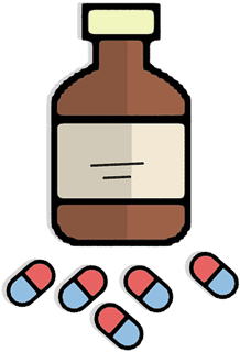
Как известно, английский язык нужен не только менеджерам, маркетологам, финансистам, фотографам, гидам, парикмахерам, но и медицинским работкам, а также людям, которые часто путешествуют. Никто не застрахован от похода в медицинскую клинику, поэтому важно хоть немного знать медицинский английский язык для того, чтобы суметь рассказать врачу о своей проблеме.
Ниже вы можете увидеть словарь медицинских терминов на английском языке:
Медицинские термины на английском
- myopia – близорукость
- farsightedness – дальнозоркость
- blindness – слепота
- running nose – насморк
- sinus trouble – гайморит
- stammer – заикание
- deafness – глухота
- otitis – отит
- dandruff – перхоть
- migraine – мигрень
- apoplexy – инсульт
- epilepsy – эпилепсия
- diabetes – диабет
- tonsillitis – тонзиллит
- sore throat – больное горло
- aphonia – потеря голоса
- disc displacement – смещение дисков
- dislocation – вывих
- cough – кашель
- bronchitis – бронхит
- heart attack – инфаркт
- fracture – перелом
- gastritis – гастрит
- ulcer – язва
- vomit – рвота
- bruise – ушиб
- insomnia – бессонница
- allergy – аллергия
- low blood pressure – низкое давление
- high blood pressure – высокое давление
- sprain – растяжение
- flu – грипп
- cold – простуда
Это лишь самые употребляемые медицинские термины на английском языке с переводом.
Названия врачей на английском

- dentist – стоматолог
- cardiologist – кардиолог
- surgeon – хирург
- traumatologist – травматолог
- dermatologist – дерматолог
- gynecologist – гинеколог
- urologist – уролог
- ophthalmologist – окулист
- therapist – физиолог
- pediatrician – педиатр
- orthopedist – ортопед
- anesthesiologist – анестезиолог
- otorhinolaryngologist – оториноларинголог
- family doctor – семейный врач
Названия медицинских инструментов
- scalpel – скальпель
- tweezers, pincers – пинцет
- needle-holder – иглодержатель
- surgical instrument – хирургический инструмент
- lancet – хирургический нож, ланцет
- drill – бормашина
- mouthwash – ополаскиватель ля полости рта
- dental floss – зубная нить
- saliva ejector – слюноотсос
- mirror – зеркало
Медицинский текст на английском языке
A doctor, Mother and Lima were sitting beside Tom’s bed. Tom was lying in bed and complaining of a headache. He had a cough, but no temperature. «What else is hurting you?» the doctor asked. «My throat and ears, and my nose won’t stop running.» «Poor little guy,» Mother said. She was worried about her son’s health. Tom sneezed and said that his stomach also hurt and he had a burning in his chest. «Very strange symptoms,» said the doctor, surprised. «I’ll prescribe aspirin, antibiotics and breathing treatments. After that you will receive several injections, and maybe even an IV.It looks like you have come down with a serious case of the flu.»
Доктор, мама и Лима сидели у постели Тома. Том лежал в кровати и жаловался на головную боль. У него был кашель, но температуры не было. «Что еще у тебя болит?» — спросил доктор. «Мое горло и уши и из носа не прекращает течь.» «Бедный малыш,» сказала мама. Она была обеспокоена здоровьем сына. Том чихнул и сказал, что живот у него тоже болит и у него жжет в груди. «Очень странные симтомы,» сказал удивленный доктор.»Я выпишу аспирин, антибиотики и ингаляции. После этого тебе сделают несколько уколов, а может, даже капельницу. Похоже, ты подхватил серьезный грипп».
Диалог с врачом на английском с переводом
Doctor: Good morning, Mr. Hocking. How are you feeling today?
Patient: I have a headache and a sore throat.
Doctor: When did it start?
Patient: About a week ago.
Doctor: Why didn’t you visit the hospital several days ago?
Patient: I thought I could cope with this by myself.
Doctor: What about other symptoms?
Patient: I also have a fever and feel pain while swallowing.
Doctor: Okay. Open your mouth and let me look at your throat. Oh, you have tonsillitis. You will need to take antibiotics. I’ll give you a prescription.
Patient: How much time should I take pills?
Doctor: Just one pill twice a day, the treatment will take up to ten days but very likely you’ll feel better in a couple of days.
Patient: Thank you.
Перевод диалога с врачом с английского на русский
Доктор: Доброе утро, мистер Хокин. Как вы чувствуете себя сегодня?
Пациент: У меня больное горло и головная боль.
Доктор: Когда это началось?
Пациент: Около недели назад.
Доктор: Почему вы не пришли в больницу несколько дней назад?
Пациент: Я думал, что справлюсь с этим самостоятельно.
Доктор: Как насчет других симптомов?
Пациент: У меня жар и я чувствую боль при глотании.
Доктор: Откройте род и дайте мне осмотреть ваше горло. О, у вас тонзиллит. Вам нужно принимать антибиотики. Я выпишу вам рецепт.
Пациент: Сколько времени мне нужно принимать лекарства?
Доктор: Одна таблетка дважды в день, лечение займет примерно десять дней, но скорее всего вам станет лучше через пару дней.
Пациент: Спасибо.
Если вы хотите усовершенствовать свой английский, в этом вам поможет lim-english.com — онлайн-самоучитель, где без репетиторов и курсов вы сможете овладеть всей необходимой информацией для того, чтобы свободно общаться на английском.
1. History of medicine
Medicine is among the most ancient of human occupations. It
began as an art and gradually developed into a science over the centuries.
There are 3 main stages in medicine development: Medicine of Ancient
Civilizations, Medicine of Middle Ages and Modern Medicine.
Early man, like the animals, was subject to illness and
death. At that time medical actions were mostly a part of ceremonial rituals.
The medicine-man practiced magic to help people who were ill or had a wound.
New civilizations, which developed from early tribes, began to study the human
body, its anatomic composition. Magic still played an important part in
treating but new practical methods were also developing. The early Indians, e.
g., set fractures and practiced aromatherapy. The Chinese were pioneers of
immunization and acupuncture. The contribution of the Greeks in medicine was
enormous. An early leader in Greek medicine was Aesculapius. His daughters,
Hygeia and Panacea gave rise to dynasties of healers (curative medicine) and
hygienists (preventive medicine). The division in curative and preventive
medicine is true today. The ethic principles of a physician were summarized by
another Greek, Hippocrates. They are known as Hippocrates Oath.
The next stage of Medicine’s development was the Middle
Ages. A very important achievement of that time was the hospital. The first
ones appeared in the 15-th century in Oriental countries and later in Europe.
Another advance of the Middle Ages was the foundation of universities during 13
– 14-th centuries. Among other disciplines students could study medicine.
During 18-th century new discoveries were made in chemistry, anatomy, biology,
others sciences. The advances of that time were invention of the stethoscope
(by Rene Laennec), vaccination for smallpox, discovery of anesthetics and
development of immunology and scientific surgery.
The next century is rise of bacteriology. Important
discoveries were made by Louis Pasteur and Robert Koch. The development of
scientific bacteriology made possible advances in surgery: using antiseptics
and control of wound infection.
Medicine in the 20-th century made enormous contribution in
the basic medical sciences. These are discovery of blood groups and vitamins,
invention of insulin and penicillin, practice of plastic surgery and
transplantation.
New words
medicine – медицина
human – человеческий
occupation – занятие
to develop – развивать
science – наука
civilization – цивилизация
Middle аges – Средние века
modern – современный
animal – животное
illness – заболевание
death – смерть
discovery – открытие
blood – кровь
2. Cell
The cell is a smallest independent unit in the body
containing all the essential properties of life. Manу types of human cells can
be grown in test tubes after beeing taken from the body. Cells which are
functionally organized are often grouped together and operate in concert as a
tissue, such as muscle tissue or nervous tissue. Various tissues may be
arranged together to form a unit called organ as the kidney, liver, heart or
lungs. Organs often function in groups called organ systems. Thus the
esophagus, stomach, раn-сreаs, liver and intestines constitute the digestive
system.
Cells are characterized by high degree of complexity and
order in both structure and function. The cell contains a number.
Of structures called cell organelles. These are responsible
for carrying out the specialized biochemical reactions characterizing each. The
many chemical reactions taking place in a cell require the establishment of
varied chemical microenvironment.
Carefully controlled transport mechanisms along with highly
effective barriers – the cell membranes – ensure that chemicals are present in
the proper region of the cell in appropriate concentration.
The cell membranes of a mixture of protein and lipid form
its surroundings.
Membranes are an essential component of almost all cells
organelles. The membrane allows only certain molecules to pass through it.
The most visible and essential organelle in a cell is the
nucleus, containing genetic material and regulating the activities of the
entire cell.
The area outside of the molecules is called the cytoplasm.
Cytoplasm contains a variety of organelles that have different functions.
New words
cell – клетка
independent – независимый
unit – единица
body – тело
all – все
lipid – жир
microenvironment – микровооружение
muscle – мышечный
nervous – нервный
digestive – пищеварительный
life – жизнь
human – человеческий
together – вместе
tissue – ткань
organ systems – системы органов
to function – функционировать
to contain – содержать
membranes – мембраны
protein – протеин
nucleus – ядро
cytoplasm – цитоплазм
different – различный
3. Tissue
A tissue is a group of cells working together to do a
special job A histologist is one who specializes in the study of tissues. The
cells, of which the tissues are made, contain from 60 to 99 % water.
Chemical reactions that are necessary for proper body function are carried on
much more readily in a water solution. The water solution and other materials
in which the tissues are bathed is slightly salty. It must be mentioned that an
insufficiency of tissues fluid is called dehydration and an abnormal
accumulation of this fluid caused a condition called edema.
Tissue classification: The 4 main groups of tissues are:
1) epithelial tissue forms elands, covers surfaces and
lines cavities;
2) connective tissue holds all parts of the body in
Place. This can be fat, cartilage, bone or blood. Blood sometimes is considered
a sort of tissue, since it contains cells and performs many of the functions of
tissues. However; the blood has many other unique characteristics;
3) nerve tissue conducts nerve impulses all over the
body;
4) the muscle tissue is designed for power-producing
contractions.
The surface of the body and of the tubes or passages leading
to the exterior and the surface of the various cavities in the body are lined
by cells which are closely approximated to each other; thus have a small amount
of intercellular substance. This lining cellular layer is called epithelium.
The nature and consistency of intercellular substance, the matrix, and the
amount and arrangement of fibers furnish the basis for the subdivision of
connective tissue into three main groups: connective tissue proper, cartilage and
bone In connective tissue the intercellular substance is soft; in cartilage it
is firm, yet flexible and elastic; in bone it is rigid due to the deposition of
calcium salt in the matrix. In multicellular organisms certain cells developed
to a high degree the properties of irritability and conductivity. These cells
form the nervous tissues.
The nervous system of higher animals is characterized by the
multiplicity of cellular forms and intercellular connections and by the
complexity of its functioning.
Muscle tissue is composed of elongated cells which have the
power of contracting or reducing their length. This property of contraction is
ultimately a molecular phenomenon and is due to the presence of protein
molecules. The following three types of muscle tissue occur in the body.
Smooth muscle tissue is found in sheet or tubes forming the
walls of many hollow or tubular organs, for example the bladder, the intes
tines of blood vessels. The cells forming this tissue are long spin dles with a
central oval nucleus.
Striated muscle tissue is composed of cylindrical fibres
often of great at length in which separate cells cannot be distinguished. Many
small nuclei are found in the fibres lie just under the surface. Cardiac muscle
resembles striated muscle in its structure, but smooth one in its action.
New words
liquid – жидкость
epithelial – эпителиальный
layer – слой
muscle – мышца
body – тело
flexible – гибкий
elastic – эластичный
nucleus – ядро
smooth – гладкий
fibre – волокно
cardiac – сердечный
4. Epidermis
The integument consists of the skin (epidermis and dermis)
and associated appendages (sweat glands, sebaceous glands, hairs, and nails)
Considered the largest body organ, the integument comprises approximately
16 % of total body weight. It is a highly specialized organ that functions
to protect the body from injury, desiccation, and infection. It also
participates in sensory reception, excretion, thermoregulation, and maintenance
of water balance.
Epidermis is the outermost layer of the integument. It is a
stratified squamous epithelial layer of ectodermal origin.
Layers of the epidermis from deep to superficial consist of
four strata. Stratum basale (stratum germinativum) is a proliferative basal
layer of columnar-like cells that contain the fibrous protein keratin. Stratum
spinosum is a multilaminar layer of cuboidal-like cells that are bound together
by means of numerous cytoplasmic extensions and desmosomal junctions.
Stratum granulosum consists of flat polygonal cells filled
with basophilic keratohyalin granules. Viewed at the electron microscopic
level, these cells also contain numerous mem brane-coating granules. Stratum
corneum is the superficial stratum of dead cells and consists of several to
many layers of flat, anucleated, and cornified (keratinized) cells. In the
epidermis of the palms and soles, a thin, transitional zone of flat
eosinophilic or pale-staining anucleated cells may occur as the stratum
lucidum. This layer is found only in regions with a thick strata corneum.
Cells of the epidermis: keratinocytes are the most numerous
and are responsible for the production of the family of keratin proteins that
provide the barrier function of the epidermis.
Melanocytes are derivatives of neural crest ectoderm. They
are found in the dermis and are also scattered among the keratinocytes in the
basal layers of the epidermis. These dendritic cells produce the pigment
melanin in the form melanosomes that are transferred to keratinocytes.
Langerhans cells are dendritic cells but are members of the
immune system and function as antigen-presenting cells. They have also been
found in other parts of the body, including the oral cavity and lymph nodes.
Merkel cells are found in the basal epidermis and appear
function in concert with nerve fibers that are closely associated with them. At
the electron microscopic level, their cytoplasm contains numerous
membrane-bound granules that resemble those of catecholamine-producing cells.
New words
epidermis – эпидермис
dermis – дерма
weight – вес
to protect – защищать
injury – рана
cytoplasmic – цитоплазматический
level – уровень
flat – плоский
palm – ладонь
thick – толстый
pigment – пигмент
melanin – меланин
nerve – нерв
5. Dermis
Dermis is a connective tissue layer of mesodermal origin
subjacent the epidermis and its basement membrane. The dermis-epidermal
junction, especially in thick skin, is characterized by numerous papillary
interdigitations of the dermal connective tissue and epidermal epithelium. This
increases the surface area of attachment and brings blood vessels in closer
proximity to the epidermal cells. The epidemis, like epithelia in general, is
devoid of blood vessel. Histologically, dermis consists of two identifiable
regions.
Papillary layer, associated principally with the dermal papillae,
is the most superficial layer. It consists of a loosely packed, irregular
meshwork of collagen fibrils that contain fine blood vessels and nerve endings.
Reticular layer is the deeper dermal layer and consists of
coarse collagen bundles intertwined with elastic fibers in a gel matrix. This
layer is a typical dense irregular connective tissue.
HYPODERMIS: this layer of loose vascular connective tissue
is infiltrated with adipocytes and corresponds to the superficial fascia of
gross anatomy. However, since it contains the deepest portions of the cutaneous
glands and hairs, it is also an important part of the skin. The hypoder-mis
fastens the skin to underlying muscles and other structures.
New words
dermis – дерма
connective – соединительный
membrane – мембрана
junction – соединение
to be characterized by – характеризоваться чем-то
numerous – значительный
to increase – увеличивать
surface – поверхность area – площадь
epidermal – эпидермальный
thick – толстый
skin – кожа
papillary – папиллярный
devoid – происходить
meshwork – ячеистая сеть
coarse – грубый
bundle – связка
interwine – сплетаться
bring – приносить
to consists of – состоять из
to contain – содержать
collagen – коллагеновый
adipocyte – жировая клетка
6. Cutaneous appendages
Cutaneous appendages are all derivatives of the epidermis.
Eccrine (merocrine) sweat glands are simple, coiled, tubular
glands that are widely distributed over the body. Secretory portions are
tightly coiled and consist of a single layer of columnar-like pyramidal cells.
Duct portions, composed of two cuboidal cell layers, are
corkscrew-shaped and open onto the epidermal surface. These glands are
important in thermal regulation.
Control of the eccrine glands is mainly by the innervation
of cholinergic fibers.
Apocrine sweat glands are also simple, coiled, tubular glands
but are much less abundant in their distribution than eccrine glands. They can
be found in the axillary, ar-eolar, and anal regions.
Secretory portions of these glands are composed of a single
layer of cuboidal or columnar cells. They are larger and have a much wider
luminal diameter than eccrine sweat glands. Myoepithelial cells surround the
secretory cells within the basement membrane and contract to facilitate
secretion.
Duct portions are similar to those of eccrine sweat glands
but open onto hair follicles instead of onto the epidermal surfaces.
Functions of these glands in humans is not at all clear.
Specialized apocrine glands in the ear canal (ceruminous glands) produce a
secretion in conjunction with adjacent sebaceous glands to form the protective
earwax (cerumen). Control of the apocrine glands is hormonal and via the
innervation of adrenergic fibers. These glands do not begin to function until
puberty.
Sebaceous glands are simple, branched holocrine aci-nar
glands. They usually discharge their secretions onto the hair shaft within hair
follicles. These glands are found in the dermis throught the skin, except on
the palms and soles.
Secretory portions consist of peripherally located,
flattened stem cells that resemble basal keratinocytes. Toward the center of
the acini, enlarged differentiated cells are engorged with lipid. Death and
fragmentation of cells nearest the duct portion result in the holocrine
mechanism of secretion.
Duct portions of sebaceous glands are composed of stratified
squamous epithelium that is continuous with the hair cat and epidermal surface.
Functions involve the lubrication of both hairs and
corni-fied layers of the skin, as well as resistance to desiccation.
Control of sebaceous glands is hormonal. Enlargement of the
acini occurs at puberty.
Hairs are long, filamentous projections consisting of dead
keratini-zed epidermal cells. Each hair derives from an epidermal invagination
called the hair follicle, which possesses a terminal hair bulb, located in the
dermis or hypo-dermis, from which the hair shaft grows. Contraction of smooth
muscles raise the hairs and dimple the epidermis («goose flesh»).
Nails, like hair, are a modified stratum corneum of the
epidermis. They contain hard keratin that forms in a manner similar to the
formation of hair. Cells continually proliferate and keratinize from the
stratum basale of the nail matrix.
New words
cutaneous – кожный
appendace – покров
tubular – трубчатый
pyramidal – пирамидальный
surface – поверхность
thermal – тепловой
innervation – иннервация
7. Matter
Matter is anything that occupies space, possesses mass and
can be perceived by our sense organs. It exists in nature in three, usually
inter convertible physical states: solids, liquids and gases. For instance,
ice, water and steam are respectively the solid, liquid and gaseous states of
water. Things in the physical world are made up of a relatively small number of
basic materials combined in various ways. The physical material of which
everything that we can see or touch is made is matter. Matter exists in three
different states: solid, liquid and gaseous. Human senses with the help of
tools allow us to determine the properties of matter. Matter can undergo a
variety of changes – physical and chemical, natural and controlled.
Chemistry and physics deal with the study of matter, its
properties, changes and transformation with energy. There are two kinds of
properties: physical – colour, taste, odour, density, hardness, solubility and
ability to conduct electricity and heat; in solids the shape of their crystals
is significant, freezing and boiling points of liquids.
Chemical properties are the changes in composition undergone
by a substance when it is subjected to various conditions. The various changes
may be physical and chemical. The physical properties are temporary. In a
chemical change the composition of the substance is changed and new products
are formed. Chemical properties are permanent.
It is useful to classify materials as solid, liquid or gas
(though water, for example, exists as solid (ice), as liquid (water) and as gas
(water vapour). The changes of state described by the terms solidify (freeze),
liquify (melt), va-pourise (evaporate) and condense are examples of physical
changes. After physical change there is still the same material. Water is water
whether it is solid, liquid or gas. Also, there is still the same mass of
material. It is usually easy to reverse a physical change.
New words
matter – материя
mass – масса
sense – чувство
organ – орган
steam – пар
to undergo – подвергать
variety – разнообрзие
change – перемена
physical – физический
chemical – химический
natural – природный
transformation – трансформация
colour – цвет
taste – вкус
odour – запах
density – плотность
hardness – твердость
solubility – растворимость
ability – возможность
to conduct – проводить
permanent – постоянный
8. Skeletal system
The components of the skeletal system are derived from
mesenchymal elements that arise from mesoderm and neural crest. Mesenchymal
cells differentiate into fibroblasts, chondroblasts, and osteoblasts, which
produce connective tissue, cartilage, and bone tissue, respectively. Bone
organs either develop directly in mesenchymal connective tissue
(intramembranous ossification) or from preformed cartilage models (endochondral
ossification) The splanch nic mesoderm gives rise to cardiac and smooth muscle.
The skeletal system develops from paraxial mesoderm. By the
end of the fourth week, the sclerotome cells form embryonic connective tissue,
known as mesenchyme. Mesenchyme cells migrate and differentiate to form
fibroblasts, chondroblasts, or osteoblasts.
Bone organs are formed by two methods.
Flat bones are formed by a process known as intra-membinous
ossification, in which bones develop directly within mesenchyme.
Long bones are formed by a process known as endochondral
ossification, in which mesenchymal cells give rise hyaline cartilage models
that subsequently become ossified
Skull formation.
Neurocranium is divided into two portions:
The membranous neurocranium consists of flat bones that
surround the brain as a vault The bones appose one another at sutures and
fontanelles, which allow overlap of bones during birth and remain membranous
until adulthood.
The cartilaginous neurocranium (chondro-cranium) of the base
of the skull is formed by fusion and ossification . -j of number of separate cartilages along the median
plate.
Viscerocranium arises primarily from the first two pharynge
arches.
Appendicular system: The pectoral and pelvic girdles and the
limbs comprise the appendicular system.
Except for the clavicle, most bones of the system are end
chondral. The limbs begin as mesenchymal buds with an apical ectodermal ridge
covering, which exerts an inductive influence over the mesenchyme.
Bone formation occurs by ossification of hyaline cartilage
models.
The cartilage that remains between the diaphysis and the epiphyses
of a long bone is known as the epiphysial plate. It is the site of growth of
long bones until they attain their final size and the epiphysial plate
disappears.
Vertebral column.
During the fourth week, sclerotome cells migrate medially to
surround the spinal cord and notochord. After proliferation of the caudal
portion of the sclerotomes, the vertebrae are formed, each consisting of the
caudal part of one sclerotome and cephalic part of the next.
While the notochord persists in the areas of the vertebral
bod ies, it degenerates between them, forming the nucleus pulposus. The latter,
together with surrounding circular fibers of the annulus fibrosis, forms the
intervertebral disc.
New words
skeletal – скелетный
mesoderm – мезодерма
cartilage – хрящ
fibroblasts – фибробласты
chondroblasts – хондробласты
osteoblasts – остеобласты
paraxial – параксиальный
flat – плоский
bone – кость
9. Muskular system
Skeletal (voluntary) system.
The dermomyotome further differentiates into the myo-tome
and the dermatome.
Cells of the myotome migrate ventrally to surround the
in-traembryonic coelom and the somatic mesoderm of the ventrolateral body wall.
These myoblasts elongate, become spindle-shaped, and fuse to form
multinucleated muscle fibers.
Myofibrils appear in the cytoplasm, and, by the third month,
cross-striations appear. Individual muscle fibers increase in diameter as
myofibrils multiply and become arranged in groups surrounded by mesenchyme.
Individual muscles form, as well as tendons that connect
muscle to bone.
Trunk musculature: By the end of the fifth week, body-wall
musculature divides into a dorsal epimere, supplied by the dorsal primary ramus
of the spinal nerve, and a ventral hypomere, supplied by the ventral primary
ramus.
Epimere muscles form the extensor muscles of the vertebral
column, and hypomere muscles give rise to lateral and ven tral flexor
musculature.
The hypomere splits into three layers. In the thorax, the
three layers form the external costal, internal intercostal, and transverse
thoracic muscle.
In the abdomen, the three layers form the external oblique,
internal oblique, and transverse abdomii muscles.
Head musculature.
The extrinsic and intrinsic muscles of the tongue are
thought to be derived from occipital myotomes that migrate forward.
The extrinsic muscles of the eye may derive from preoptic
myotomes that originally surround the prochordal plate.
The muscles of mastication, facial expression, the pharynx,
and the larynx are derived from different pharyngeal arches and maintain their
innervation by the nerve of the arch of origin.
Limb musculature originates in the seventh week from soma
mesoderm that migrates into the limb bud. With time, the limb musculature splits
into ventral flexor and dorsal extern groups.
The limb is innervated by spinal nerves, which penetrate the
limb bud mesodermal condensations. Segmental branches of the spinal nerves fuse
to form large dorsal a ventral nerves.
The cutaneous innervation of the limbs is also derived from
spinal nerves and reflects the level at which the limbs arise.
Smooth muscle: the smooth muscle coats of the gut, trachtea,
bronchi, and blood vessels of the associated mesenteries are derived from
splanchnic mesoderm surrounding the gastrointestinal tract. Vessels elsewhere
in the body obtain their coat from local mesenchyme.
Cardiac muscle, like smooth muscle, is derived from
splanchnic mesoderm.
New words
ventral – брюшной
somatic – соматический
cytoplasm – цитоплазма
cross-striations – поперечные бороздчатости
extensor – разгибающая мышца
dorsal – спинной
ivertebral – позвоночный
arche – дуга
abdomen – живот
facial – лицевой
branch – ветвь
10. Skeleton
The bones of our body make up a skeleton. The skeleton forms
about 18 % of the weight of the human body.
The skeleton of the trunk mainly consists of spinal column
made of a number of bony segments called vertebrae to which the head, the
thoracic cavity and the pelvic bones are connected. The spinal column consists
of 26 spinal column bones.
The human vertebrae are divided into differentiated groups.
The seven most superior of them are the vertebrae called the cervical
vertebrae. The first cervical vertebra is the atlas. The second vertebra is
called the axis.
Inferior to the cervical vertebrae are twelve thoracic
vertebrae. There is one rib connected to each thoracic vertebrae, making 12
pairs of ribs. Most of the rib pairs come together ventrally and join a flat
bone called the sternum.
The first pairs or ribs are short. All seven pairs join the
sternum directly and are sometimes called the «true ribs». Pairs 8, 9, 10 are
«false ribs». The eleventh and twelfth pairs of ribs are the «floating ribs».
Inferior to the thoracic vertebrae are five lumbar
vertebrae. The lumbar vertebrae are the largest and the heaviest of the spinal
column. Inferior to the lumbar vertebrae are five sacral vertebrae forming a
strong bone in adults. The most inferior group of vertebrae are four small
vertebrae forming together the соссуx.
The vertebral column is not made up of bone alone. It also
has cartilages.
New words
skeleton – скелет
make up – составлять
weight – вес
trunk – туловище
vertebrae – позвоночник
thoracic cavity – грудная клетка
pelvic – тазовый
cervical – шейный
atlas – 1 шейный позвонок
sternum – грудина
mainly – главным образом
axis – ось
spinal column – позвоночник
inferior – нижний
rib – ребро
pair – пара
sacral – сакральный
соссуx – копчик
floating – плавающий
forming – формирующий
cartilage – хрящ
lumbar – поясничный
adult – взрослый
11. Muscles
Muscles are the active part of the motor apparatus; their
contraction produces various movements.
The muscles may be divided from a physiological standpoint
into two classes: the voluntary muscles, which are under the control of the
will, and the involuntary muscles, which are not.
All muscular tissues are controlled by the nervous system.
When muscular tissue is examined under the microscope, it is
seen to be made up of small, elongated threadlike cells, which arc called
muscle fibres, and which are bound into bundles by connective tissue.
There are three varieties of muscle fibres:
1) striated muscle fibres, which occur in voluntary
muscles;
2) unstriated muscles which bring about movements in
the internal organs;
3) cardiac or heart fibres, which are striated like
(1), but are otherwise different.
Muscle consists of threads, or muscle fibers, supported by
connective tissue, which act by fiber contraction. There are two types of
muscles smooth and striated. Smooth, muscles are found in the walls of all the
hollow organs and tubes of the body, such as blood vessels and intestines.
These react slowly to stimuli from the autonomic nervous system. The striated,
muscles of the body mostly attach to the bones and move the skeleton. Under the
microscope their fibres have a cross – striped appearance. Striated muscle is
capable of fast contractions. The heart wall is made up of special type of
striated muscle fibres called cardiac muscle. The body is composed of about 600
skeletal muscles. In the adult about 35–40 % of the body weight is formed
by the muscles. According to the basic part of the skeleton all the muscles are
divided into the muscles of the trunk, head and extremities.
According to the form all the muscles are traditionally
divided into three basic groups: long, short and wide muscles. Long muscles
compose the free parts of the extremities. The wide muscles form the walls of
the body cavities. Some short muscles, of which stapedus is the smallest muscle
in the human body, form facial musculature.
Some muscles are called according to the structure of their
fibres, for example radiated muscles; others according to their uses, for
example extensors or according to their directions, for example, –
oblique.
Great research work was carried out by many scientists to
determine the functions of the muscles. Their work helped to establish that the
muscles were the active agents of motion and contraction.
New words
muscles – мышцы
active – активный
motor apparatus – двигательный аппарат
various – различный
movement – движение
elongated – удлиненный
threadlike – нитевидный
be bound – быть связанным
ability – возможность
capable – способность
scientist – ученый
basic – основной
12. Bones
Bone is the type of connective tissue that forms the body s
supporting framework, the skeleton. Serve to protect the internal organs from
injury. The bone marrow inside the bones is the body’s major producer of both
red and white blood cells.
The bones of women are generally lighter than those of men,
while children’s bones are more resilient than those of adults Bones also respond
to certain physical physiological changes: atrophy, or waste away.
Bones are generally classified in two ways. When classified
on the basis of their shape, they fall into four categories: flat bones, such
as the ribs; long bones, such as the thigh bone; short bones, such as the wrist
bones; and irregular bones, such as the vertebrae. When classified on the basis
of how they develop, bones are divided into two groups: endochondral bones and
intramembraneous bones. Endochondral bones, such as the long bones and the
bones at the base of the skull, develop from cartilage tissue Intramembraneous
bones, such as the flat bones of the roof of the skull, are not formed from
cartilage but develop under or within a connective tissue membrane. Although
endochondral bones and intramembraneous bones form in different ways, they have
the same structure.
The formation of bone tissue (ossification) begins early in
embryological development. The bones reach their full size when the person is
about 25.
Most adult bone is composed of two types of tissue: anouter
layer of compact bone and an inner layer of spongy bone. Compact bone is strong
and dense. Spongy bone is light and porous and contains bone marrow The amount
of each type of tissue varies in different bones. The flat bones of the skull
consist almost entirely of compact bone, with very little spongy tissue. In a
long bone, such as the thigh bone, the shaft, called the diaphysis, is made up
largely of compact bone. While the ends, called epyphyses, consist mostly of spongy
bone. In a long bone, marrow is also present inside the shaft, in a cavity
called the medullary cavity.
Surrounding every bone, except at the surface where it meets
another bone, is a fibrous membrane called the periosteum. The outer layer of
the periosteum consists of a network of densely packed collagen fibres and
blood vessels. This layer serves for the attachment of tendons, ligaments, and
muscles to the bone and is also important in bone repair.
The inner layer of the periosteum has many fibres, called
fibres of Sharpey, which penetrate the bone tissue, anchoring the periosteum to
the bone. The inner layer also has many bone-forming cells, or osteoblasts,
which are responsible for the bone’s growth in diameter and the production of
new bone tissue in cases of fracture, infection.
In addition to the periosteum, all bones have another
membrane, the endosteum. It lines the marrow cavity as well as the smaller
cavities within the bone. This membrane, like the inner layer of the
periosteum, contains os-teoblasts, and is important in the formation of new
bone tissue.
13. Bones. Chemical structure,
Bone tissue consists largely of a hard substance called the
matrix. Embedded in the matrix are the bone cells, or osteocytes. Bone matrix
consists of both organic and inorganic materials. The organic portion is made
up chiefly of collagen fibres. The inorganic portion of matrix constitutes
about two thirds of a bone’s total weight. The chief inorganic substance is
calcium phosphate, which is responsible for the bone’s hardness. If the organic
portion were burned out the bone would crumble under the slightest pressure. In
the formation of intramembraneous bone, certain cells of the embryonic
connective tissue congregate in the area where the bone is to form. Small blood
vessels soon invade the area, and the cells, which have clustered in strands,
undergo certain changes to become osteoblasts. The cells then begin secreting
collagen fibers and an intercellular substance. This substance, together with
the collagen fibers and the connective tissue fibers already present, is called
osteoid. Osteoid is very soft and flexible, but as mineral salts are deposited
it becomes hard matrix. The formation of endochondral bone is preceded by the
formation of a cartilaginous structure similar in shape to the resulting bone.
In a long bone, ossification begins in the area that becomes the center of the
shaft. In this area, cartilage cells become osteoblasts and start forming bone
tissue This process spreads toward either end of the bone. The only areas where
cartilage is not soon replaced by bone tissue are the regions where the shaft
joins the two epiphyses. These areas, called epiphyseal pla-res, are
responsible for the bones continuing growth in length. The bone’s growth in
diameter is due to the addition of layers of bone around the outside of the
shaft As they are formed, layers of bone on the inside of the shaft are
removed. In all bones, the matrix is arranged in layers called lamellae. In
compact bone, the lamellae are arranged concentrically around blood vessels,
and the space containing each blood vessel is called a Haver-sian canal. The
osteocytes are located between the lamellae, and the canaliculi containing
their cellular extensions connect with the Haversian canals, allowing the
passage of nutrients and other materials between the cells and the blood
vessels. Bone tissue contains also many smaller blood vessels that extend from
the periosteum and enter the bone through small openings. In long bones there
is an additional blood supply, the nutrient artery, which represents the chief
blood supply to the marrow. The structure of spongy is similar to that of
compact bone. However, there are fewer Haversian canals, and the lamellae are
arranged in a less regular fashion, forming spicules and strands known as
trabeculae.
New words
bone – кость
internal – внешний
phosphorus – фосфор
atrophy – атрофия
spongy – губчатый
tendon – сухожилие
ligament – связка
flexible – гибкий
periosteum – надкостница
osteoblast – остеобласт (клетка, образующая кость)
rigidity – неподвижность
shape – форма
to crumble – крошиться
to congregate – собираться
epiphyseal – относящийся к эпифизу
shaft – ствол, тело (длинной) кости, диафиз
14. Skull
Bones of the skull: the neurocranium (the portion of the
skull that surrounds and protects the brain) or the viscerocra-nium (i. e., the
skeleton of the face). Bones of the neurocranium Frontal, Parietal, Temporal,
Occipital, Ethmoid, Sphenoid.
Bones of the viscerocranium (surface): Maxilla, Nasal,
Zygomatic, Mandible. Bones of the viscerocranium (deep): Ethmoid, Sphenoid,
Vomer, Lacrimal, Palatine, Inferior nasal concha. Articulations: Most skull
bones meet at immovable joints called sutures. The coronal suture is between
the frontal and the parietal bones The sagittal suture is between two parietal
bones. The lambdoid suture is between the parietal and the occipital bones. The
bregma is the point at which the coronal suture intersects the sagittal suture
The lambda is the point at which the sagittal suture
intersects the lambdoid suture. The pterion is the point on the lateral aspect
of the skull where the greater wing of the sphenoid, parietal, frontal, and
temporal bones converge. The temporomandibular joint is between the mandibular
fossa of the temporal bone and the condylar process of the mandible.
The parotid gland is the largest of the salivary glands.
Structures found within the substance of this gland include the following:
Motor branches of the facial nerve CN VII enters the parotid gland after
emerging from the stylomastoid foramen at the base of the skull. Superficial
temporal artery and vein. The artery is a terminal branch of the external
carotid artery.
Retromandibular vein, which is formed from the maxillary and
superficial temporal veins.
Great auricular nerve, which is a cutaneous branch of the
cer vical plexus. Auriculotemporal nerve, which is a sensory branch of V3. It
supplies the TMJ and conveys postganglionic parasympathetic fibers from the
otic ganglion to the parotid gland Parotid (Stensen s) duct, which enters the
oral cavity at the level of the maxillary second molar. The facial artery
is a branch of the external carotid artery in the neck. It
terminates as the angular artery near the bridge of the
nose.
The muscles of face
New words
brain – мозг
frontal – лобная
parietal – теменная
temporal – височная
occipital – затылочная
ethmoid – решетчатая
maxilla – верхняя челюсть
zygomatic – скуловой
mandible – нижняя челюсть
sphenoid – клиновидная
vomer – сошник
lacrimal – слезная
palatine – небная
nasal concha – носовая раковина
15. Neck. Cervical vertebrae, cartilages, triangels
Cervical vertebrae: There are seven cervical vertebrae of
which the first two are atypical. All cervical vertebrae have the foramina
transversaria which produce a canal that transmits the vertbral artery and
vein.
Atlas: This is the first cervical vertebra (C1). It has no
body and leaves a space to accommodate the dens of the second cervical
vertebra. Axis: This is the second cervical vertebra (C2). It has odontoid
process, which articulates with the atlas as a pivot joint. Hyoid bone is a
small U-shaped bone, which is suspended by muscles and ligaments at the level
of vertebra C3.
Laryngeal prominence is formed by the lamina of the thyroid
cartilage.
Cricoid cartilage. The arch of the cricoid is palpable below
the thyroid cartilage and superior to the first tracheal ring (vertebral level
C6). Triangles of the neck: The neck is divided into a posterior and an
arterior triangle by the sternocleidomastoid muscle. These triangles are
subdivided by smaller muscles into six smaller triangles. Posterior triangle is
bound by the sternocleidomastoid, the clavicle, and the trapezius. Occipital
triangle is located above the inferior belly of the omohyoid muscle. Its
contents include the following: CN XI Cutaneous branches of the cervical plexus
are the lesser occipital, great auricular, transverse cervical, and
supaclavicular nerves.
Subclavian (omoclavicular, supraclavicular) triangle is
located below the inferior belly of the omohyoid. Its contents include the
following: Brachial plexus supraclavicular portion The branches include the
dorsal scapular, long thoracic, subclavius, and suprascapular nerves.
The third part of the subclavian artery enters the
subclavian triangle.
The subclavian vein passes superficial to scalenus anterior
muscle. It receives the external jugular vein.
Anterior triangle is bound by the sternocleidomastoid muse
the midline of the neck, and the inferior border of the body of the mandible.
Muscular triangle is bound by the sternocleidomastoid muscle, the superior
belly of the omohyoid muscle, and the midline of the neck. Carotid (vascular)
triangle is bound by the sternocleidomastoid muscle, the superior belly of the
omohyoid muscle and the posterior belly of the digastric muscle. The carotid
triangle contains the following: Internal jugular vein; Common carotid artery,
bifurcates and form the internal and external carotid arteries. The external
carotid artery has six branches (i. e., the superior thyroid; the ascending
pharyngeal, the lingual, the facial, the occipital, and the posterior auricular
arteries). Vagus nerve; hypoglossal nerve; internal and external laryngeal
branches of the superior laryngeal branch of the vagus nerve. Digastric
(sub-mandibular) triangle is bound by the anterior and posterior bellies of the
digastric muscle and the inferi or border of the body of the mandible. It
contains the submandibular salivary gland. Submental triangle is bound by the
anterior belly of the digastric muscle, the hyoid bone, and the midline of the
neck. It contains the submental lymph nodes.
16. Neck. Root, fascies of the neck
Root of neck: This area communicates with the superior medi
astinum through the thoracic inlet. Structures of the region include the
following: subclavian artery and vein. The subclavian artery passes poste rior
to the scalenus anterior muscle, and the vein passes ante rior to it Branches
of the artery include: vertebral artery; thyrocervical trunk, which gives rise
to the inferior thyroid, the transverse cervical, and the suprascapular
arteries; Internal thoracic artery.
Phrenic nerve is a branch of the cervical plexus, which
arises from C3, C4, and C5. It is the sole motor nerve to the diaphragm. It
crosses the anterior scalene muscle from lateral to medial to enter the
thoracic inlet.
Recurrent laryngeal nerve is a branch of the vagus nerve.
This mixed nerve conveys sensory information from the laryngeal; mucosa below
the level of the vocal folds and provides motor innervation to all the
intrinsic muscles of the larynx except the cricothyroid muscle.
Thoracic duct terminates at the junction of the left
subclavian and the left internal jugular veins On the right side of the body,
the right lymphatic duct terminates in a similar fashion.
Fascias of the neck Superficial investing fascia encloses
the platysma, a muscle of facial expression, which has migrated to the neck
Deep investing fascia surrounds the trapezius and
ster-noclei – domastoid muscles.
Retropharyngeal (visceral) fascia surrounds the pharynx.
Prevertebral fascia invests the prevertebral muscles of the
nee (i. e., longus colli, longus capitis) This layer gives rise to a derivative
known as the alar fascia.
The major muscle groups and their innervations. A simple
method of organizing the muscles of the neck is based on two basic principles:
(1) The muscles may be arranged in group according to their functions; and (2)
all muscles in a group share common innervation with one exception in each
group.
Group 1: Muscles of the tongue. All intrinsic muscles plus
all but one of the extrinsic muscles (i. e., those containing the suffix,
glossus) of the tongue are supplied by CN XII. The one exception is
palatoglossus, which is supplied by CN X.
Group 2: Muscles of the larynx. All but one of the intrinsic
muscles of the larynx are supplied by the recurrent la-ryngeal branch of the vagus
nerve. The sole exception is the cricothyroid muscle, which is supplied by the
external laryngeal branch of the vagus.
Group 3: Muscles of the pharynx. All but one of the
longitudinal and circular muscles of the pharynx are supplied by CNs X and XI (cranial
portion). The sole exception is the stylopharyngeus muscle, which is supplied
by CN IX.
Group 4: Muscles of the soft palate. All but one of the
muscles of the palate are supplied by CNs X and XI (cranial portion). The sole
exception is the tensor veli palatini, which is supplied CN V3.
Group 5: Infrahyoid muscles. All but one of the infrahyo-id
muscles are supplied by the ansa cervicalis of the cervical olexus (C1, C2, and
C3). The exception is the thy-rohyoid, which is supplied by a branch of C1.
(This branch of C1 also supplies the geniohyoid muscle).
New words
neck – шея
cervical – цервикальный
vertebrae – позвоночник
cricoid cartilage – перстневидный хрящ гортани
scapulae – лопатка
scalene – лестничная мышца
brachial plexus – плечевое сплетение
vagus nerve – блуждающий нерв
hypoglossal nerve – подъязычный нерв
laryngeal branches – гортанные ветви
17. Thoracic wall
There are 12 thoracic vertebrae. Each rib articulates with
the body of the numerically corresponding vertebra and the one below it.
Sternum: the manubrium articulates with the clavicle and the first rib. It
meets the body of the sternum at the sternal angel an important clinical
landmark.
The body articulates directly with ribs 2–7; it articulates
interiorly with the xiphoid process.
Ribs and costal cartilages: there are 12 pairs of ribs,
which are attached posteriorly to thoracic vertebrae.
Ribs 1–7 attach directly to the sternum by costal
cartilages.
Ribs 8 – 10 attach to the costal cartilage of the rib above.
Ribs 11 and 12 have no anterior attachments. The costal groove is located along
the inferior border of each rib and provides protection for the intercostal
nerve artery, and vein.
There are 11 pairs of external intercostal muscles.
These muscles fill the intercostal spaces from the tubercles
of ribs posteriorly to the costochondral junctions anteriorly. There are 11
pairs of internal intercostal muscles.
These muscles fill the intercostal spaces anteriorly from
the sternum to the angles of the ribs posteriorly.
Innermost intercostal muscles: the deep layers of the internal
intercostal muscles are the innermost intercostal muscles.
Subcostalis portion: Fibers extend from the inner surface of
the angle of one rib to the rib that is inferior to it.
Internal thoracic vessels, branches of the subclavian
arteries, run anterior to these fibers. Intercostal structures
Intercostal nerves: there are 12 pairs of thoracic nerves,
11 intercostal pairs, and 1 subcostal pair.
Intercostal nerves are the ventral primary rami of thoracic
spinal nerves. These nerves supply the skin and musculature of the thoracic and
abdominal walls.
Intercostal arteries: there are 12 pairs of posterior and
anterior arteries, 11 intercostal pairs, and 1 subcostal pair.
Anterior intercostal arteries.
Pairs 1–6 are derived from the internal thoracic arteries.
Pairs 7–9 are derived from the musculophrenic arteries.
Posterior intercostal arteries: the first two pairs arise
from the superior intercostal artery, a branch of the costo-cervical trunk of
the subcla vian artery.
Nine pairs of intercostal and one pair of subcostal arter
ies arise from the thoracic aorta.
Intercostal veins: Anterior branches of the intercostal
veins drain to the internal thoracic and musculophrenic veins.
Posterior branches drain to the azygos system of veins.
Lymphatic drainage of intercostal spaces: anterior drainage
is to the internal thoracic (parasternal) nodes.
Posterior drainage is to the paraaortic nodes of the poste
rior mediastinum.
New words
thoracic – грудной
wall – стенка
clavicle – ключица
xiphisternal – грудинный
groove – углубление
intercostal – межреберный
subcostal – подкостный
transversus – поперечный
musculophrenic – мышечный грудобрюшной
paraaortic – парааортальный
mediastinum – средостение
18. Blood. Formed elements of the blood. Erythrocytes and platelets
Blood is considered a modified type of connective tissue
Mesodermal is composed of cells and cell frag ments (erythrocytes, leukocytes,
platelets), fibrous proteins (fibrinogen), and an extracellular fluid and
proteins (plasma). It also contains cellular elements of the immune system as
well as humoral factors
The formed elements of the blood include erythrocytes,
leukocytes, and platelets.
Erythrocytes, or red blood cells, are important in trans
porting oxygen from the lungs to tissues and in returning carbon dioxide to the
lungs. Oxygen and carbon dioxide carried in the RBC combine with hemoglobin to
form oxyhemoglobin and carbaminohemoglobin, respectively.
Mature erythrocytes are denucleated, biconcave disks with a
diameter of 7–8 mm. The biconcave shape results in a 20–30 % increase in
sur face area compared to a sphere.
Erythrocytes have a very large surface area: volume ratio
that allows for efficient gas transfer. Erythrocyte membranes are remarkably
pliable, enabling the cells to squeeze through the narrowest capillaries In
sickle cell anemia, this plasticity is lost, and the subsequent clogging of
capillaries leads to sickle crisis. The normal concentration of erythrocytes in
blood is 3,5–5,5 million/mm3 in women and 4,3–5,9 million/mm3in
men. The packed volume of blood cells per total volume of known as the
hematocrit. Normal hematocrit values are 46 % for women and 41–53 %
for men.
When aging RBCs develop subtle changes, macrophages in the
bone marrow, spleen, and liver engulf and digest them. The iron is carried by
transferring in the blood to certain tissues, where it combines with
apoferritin to form ferritin. The heme is catabolized into biliver-din, which
is converted to bilirubin. The latter is secreted with bile salts.
Platelets (thromboplastids) are 2–3 mm in diameter.
They are a nuclear, membrane-bound cellular fragments
derived by cytoplasmic fragmentation of giant cells, called megakaryocytes, in
the bone marrow.
They have a short life span of approximately 10 days.
There are normally 150 000–400 000 platelets per mm3 of
blood. Ultrastructurally, platelets contain two portions: a peripheral,
light-staining hyalomere that sends out fine cytoplasmic processes, and a
central, dark-staining granulomere that con tains mitochondria, vacuoles, glycogen
granules, and granules. Platelets seal minute breaks in blood vessels and
maintain endothelial integrity by adhering to the damaged vessel in a process
known as platelet aggregation. Platelets are able to form a plug at the rupture
site of a vessel because their mem brane permits them to agglutinate and adhere
to surfaces.
Platelets aggregate to set up the cascade of enzymatic reac
tions that convert fibrinogen into the fibrin fibers that make up the clot.
New words
mesodermal – мезодермальный
erythrocytes – эритроциты
platelets – тромбоциты
carbon – углерод
dioxid – диоксид
span – промежуток
light-staining – легкое окрашивание
to aggregate – соединяться
19. Blood. Formed elements of the blood. Leukocytes
Leukocytes, or white blood cells, are primarily with the
cellular and humoral defense of the organism foreign materials. Leukocytes are
classified as granulocytes (neutrophils, eosinophils, basophils) and
agranulocytes (lympmonocytes).
Granulocytes are named according to the staining properties
of their specific granules. Neutrophils sare 10–16 mm in diameter.
They have 3–5 nuclear lobes and contain azurophilic granules
(lysosomes), which contain hydrolytic enzymes for bacterial destruction, in
their cytoplasm. Neutrophils are phagocytes that are drawn (chemotaxis) to
bacterial chemoattractants. They are the primary cells involved in the acute
inflammatory response and represent 54–62 % of leukocytes.
Eosinophils: they have a bilobed nucleus and possess acid
granulations in their cytoplasm. These granules contain hydrolytic enzymes and
peroxidase, which a discharged into phagocytic vacuoles.
Eosinophils are more numerous in the blood during allergic
diseases; they norma asent only – 3 % of leukocytes.
Basophils: they possess large spheroid granules, which are
basophilic and metachromatic
Basophils degranulate in certain immune reaction, releasing
heparin and histamine into their surroundings They also release additional
vasoactive amines and slow reacting substance of anaphylaxis (SRS-A) consisting
of leu-kotrienes LTC4, LTD4, and LTE4. They represent less than 1 % – of
leukocytes
Agranulocytes are named according to their lack of specific
granules. Lymphocytes are generally small cells measuring 7 – 10 mm in diameter
and constitute 25–33 % of , leukocytes. They con tain circular
dark-stained nuclei and scanty clear blue cyto plasm. Circulating lymphocytes
enter the blood from the lymphatic tissues. Two principal types of
immunocompetent lymphocytes can be identified T lymphocytes and В lymphocytes.
T cells differentiate in the thymus and then circulate in
the peripheral blood, where they are the principal effec tors of cell-mediated
immunity. They also function as helper and suppressor cells, by modulating the
immune response through their effect on В cells, plasma cells, macrophages, and
other T Cells.
В cells differentiate in bone marrow. Once activated by
contact with an antigen, they differentiate into plasma cells, which synthesize
antibodies that are secreted into the blood, intercellular fluid, and lymph. В
lymphocytes also give rise to memory cells, which differentiate into plas ma
cells only after the second exposure to the antigen. Monocytes vary in diameter
from 15–18 mm and are the largest of the peripheral blood cells. They
constitute 3–7 % of leukocytes.
Monocytes possess an eccentric nucleus. The cytoplasm has a
ground-glass appearance and fine azurophilic granules.
Monocytes are the precursors for members of the mo-nonuclear
phagocyte system, including tissue macrophages (histiocytes), osteoclasts,
alveolar macrophages, and Kupffer cells of the liver.
New words
mesodermal – мезодермальный
erythrocytes – эритроциты
leukocytes – лейкоциты
fibrous proteins – волокнистые белки
immune – иммунный
humoral – гуморальный
to contain – содержать
nuclei – ядра
20. Plasma
Plasma is the extracellular component of blood. It is an
aqueous solution containing proteins, inorganic salts, and organic com pounds
Albumin is the major plasma protein that maintains the osmotic pressure of
blood. Other plasma proteins include the globulins (alpha, beta, gamma) and
fibrinogen, which is necessary for the formation of fibrin in the final step of
blood coagulation Plasma is in equilibrium with tissue interstitial fluid
through capil lary walls; therefore, the composition of plasma may be used to
judge the mean composition of the extracellular fluids Large blood proteins
remain in the intravascular compartment and do not equilibrate with the
interstitial fluid Serum is a clear yellow fluid that is separated from the
coagulum during the process of blood clot formation. It has the same com
position as plasma, but lacks the clotting factors (especially fib rinogen).
Lymphatic vessels
Lymphatic vessels consist of a, fine network of thin-walled
vessels that drain into progressively larger and progressively thicker-walled
collecting trunks. These ultimately drain, via the thoracic duct and right
lymphatic duct, into the left and right subclavian veins at their angles of
junction with the internal jugular veins, respectively. The lymphatics serve as
a one-way (i. e., toward the heart) drainage sys tem for the return of tissue
fluid and other diffusible substances, including plasma proteins, which
constantly escape from the blood through capillaries. They are also important
in serving as a conduit for channeling lymphocytes and antibodies produced in
lymph nodes into the blood circulation.
Lymphatic capillaries consist of vessels lined with
endothelial cells, which begin as blind-ended tubules or saccules in most tis
sues of the body Endothelium is attenuated and usually lacks a continuous basal
lamina. . Lymphatic vessels of large diameter resemble veins in their struc
ture but lack a clear-cut separation between layers. Valves are more numerous
in lymphatic vessels. Smooth muscle cells in the media layer engage in rhythmic
contraction, pumping lymph toward the venous system. Smooth muscle is
well-developed in large lymphatic ducts.
Circulation of lymph is slower than that of blood, but it is
nonetheless an essential process. It has been estimated that in a single day,
50 % or more of the total circulating protein leaves the blood circulation
at the capillary level and is recaptured by the lymphatics.
Distribution of lymphatics is ubiquitous with some notable
excep tions, including epithelium, cartilage, bone, central nervous sys tem,
and thymus.
New words
plasma – плазма
extracellular – внеклеточный
aqueous – водный
solution – раствор
proteins – белки
inorganic – неорганический
salts – соли
organic – органический
albumin – альбумин
globulins – глобулины
alpha – альфа
beta – бета
gamma – гамма
fibrinogen – фибриноген
lymphatic – лимфатический
vessel – сосуд
endothelium – эндотелий
circulation – кровообращение
lymph – лимфа
ubiquitous – вездесущий
notable – известный
21. Hematopoietic tissue. Erythropoiesis
Hematopoietic tissue is composed of reticular fibers and
cells, blood vessels, and sinusoids (thin-walled blood channels). Myeloid, or
blood cell-forming tissue, is found in the bone marrow and provides the stem
cells that develop into erythrocytes, granulocytes, agranulocytes, and
platelets. Red marrow is characterized by active hemato-poiesis; yellow bone
marrow is inactive and contains mostly fat cells. In the human adult,
hematopoiesis takes place in the mar row of the flat bones of the skull, ribs
and sternum, the vertebral column, the pelvis, and the proximal ends of some
long bones. Erythropoiesis is the process of RBC formation. Bone marrow stem
cells (colony-forming units, CFUs) differentiate into proerythroblasts under
the influence of the glycoprotein erythropoietin, which is produced by the
kidney.
Proerythroblast is a large basophilic cell containing a
large spherical euchromatic nucleus with prominent nucleoli.
Basophilic erythroblast is a strongly basophilic cell with
nucleus that comprises approximately 75 % of its mass. Numerous
cytoplasmic polyribosomes, condensed chro-matin, no visible nucleoli, and
continued hemoglobin synthesis characteristics of this cell.
Polychromatophilic erythroblast is the last cell in this
line undergoes mitotic divisions. Its nucleus comprises approximately 50 %
of its mass and contains condensed chroma-tin which appears in a «checkerboard»
pattern. The po-lychnsia of the cytoplasm is due to the increased quantity of
acidophilic hemoglobin combined with the basophilia of cytoplasmic
polyribosomes.
Normoblast (orthochromatophilic erythroblast) is a cell with
a small heterochromatic nucleus that comprises approximately 25 % of its
mass. It contains acidophilic cytoplasm because the large amount of hemoglobin
and degenerating organelles. The pyknotic nucleus, which is no longer capable
of division, is extruded from the cell.
Reticulocyte (polychromatophilic erythrocyte) is an immature
acidophilic denucleated RBC, which still contains some ribosomes and
mitochondria involved in the synthesis of a small quantity of hemoglobin.
Approximately 1 % of the circulating RBCs are reticulocytes.
Erythrocyte is the mature acidophilic and denucleated RBC.
Erythrocytes remain in the circulation approximately 120 days and are then
recycled by the spleen, liver, and bone marrow.
New words
reticular – сетчатый
sinusoids – синусоиды
granulocytes – гранулоциты
agranulocytes – агранулоциты
active – активный
yellow – желтый
glycoprotein – гликопротеин
erythropoietin – эритропоэтин
amount – количество
hemoglobin – гемоглобин
degenerating – дегенерирующие
condensed – сжатый
22. Hematopoietic tissue. Granulopoiesis, thrombopoiesis
Granulopoiesis is the process of granulocyte formation. Bone
marrow stem cells differentiate into all three types of granulocytes.
Myeloblast is a cell that has a large spherical nucleus
containing delicate euchromatin and several nucleoli. It has a basophilic
cytoplasm and no granules. Myeloblasts divide differentiate to form smaller
promyelocytes.
Promyelocyte is a cell that contains a large spherical
indented nucleus with coarse condensed chromatin. The cytoplasm is basophilic
and contains peripheral azurophi-lic granules.
Myelocyte is the last cell in this series capable of
division. The nucleus becomes increasingly heterochromatic with subsequent
divisions. Specific granules arise from the Golgi apparatus, resulting in
neutrophilic, eosinophilic, and basophilic myelocytes.
Metamyelocyte is a cell whose indented nucleus exhibits lobe
formation that is characteristic of the neutrophil, eos-inophil, or basophil.
The cytoplasm contains azurophilic granules and increasing numbers of specific
granules. This cell does not divide. Granulocytes are the definitive cells that
enter the blood. Neutrophilic granulocytes exhibit an intermediate stage called
the band neutrophil. This is the first cell of this series to appear in the
peripheral blood.
It has a nucleus shaped like a curved rod or band.
Bands normally constitute 0,5–2 % of peripheral WBCs;
they subsequently mature into definitive neutrophils.
Agranulopoiesis is the process of lymphocyte and mono-cyte
for mation. Lymphocytes develop from bone marrow stem cells (lymphoblasts).
Cells develop in bone marrow and seed the secondary lymphoid organs (e. g., tonsils,
lymph nodes, spleen). Stem cells for T cells come from bone marrow, develop in
the thymus and, subsequently, seed the secondary lym phoid organs.
Promonocytes differentiate from bone marrow stem cells
(monoblasts) and multiply to give rise to monocytes.
Monocytes spend only a short period of time in the marrow
before being released into the bloodstream.
Monocytes are transported in the blood but are also found in
connective tissues, body cavities and organs.
Outside the blood vessel wall, they are transformed into
macrophages of the mononuclear phagocyte system.
Thrombopoiesis, or the formation of platelets, occurs in the
red bone marrow.
Megakaryoblast is a large basophilic cell that contains a
U-shaped or ovoid nucleus with prominent nucleoli. It is the last cell that
undergoes mitosis.
Megakaryocytes are the largest of bone marrow cells, with
diameters of 50 mm or greater. They undergo 4–5 nuclear divi sions without
concomitant cytopla-smic division. As a result, the megakaryocyte is a cell
with polylobulated, polyploid nucleus and abundant granules in its cytoplasm.
As megakaryocyte maturation proceeds, «curtains» of platelet demarcation
vesicles form in the cytoplasm. These vesicles coalesce, become tubular, and
eventually form platelet demarcation membranes. These membranes fuse to give
rise to the membranes of the platelets.
A single megakaryocyte can shed (i. e., produce) up to 3,500
platelets.
New words
capable – способный
spherical – сферический
indented – зазубренный
chromatin – хроматин
23. Arteries
Arteries are classified according to their size, the
appearance of their tunica media, or their major function.
Large elastic conducting arteries include the aorta and its
large branches. Unstained, they appear yellow due to their high con tent of
elastin.
The tunica intima is composed of endothelium and a thin sub
jacent connective tissue layer. An internal elastic membrane marks the boundary
between the intima and media.
The tunica media is extremely thick in large arteries and
con sists of circularly organized, fenestrated sheets of elastic tissue with
interspersed smooth muscle cells. These cells are responsi ble for producing
elastin and other extracellular matrix com ponents. The outermost elastin sheet
is considered as the external elastic membrane, which marks the boundary
between the media and the tunica adventitia.
The tunica adventitia is a longitudinally oriented
collection of collagenous bundles and delicate elastic fibers with associated
fibroblasts. Large blood vessels have their own blood supply (vasa vasorum),
which consists of small vessels that branch profusely in the walls of larger
arteries and veins. Muscular distributing arteries are medium-sized vessels
that are characterized by their predominance of circularly arranged smooth
muscle cells in the media interspersed with a few elastin compo nents. Up to 40
layers of smooth muscle may occur. Both internal and external elastic limiting
membranes are clearly demonstrated. The intima is thinner than that of the
large arteries.
Arterioles are the smallest components of the arterial tree.
Generally, any artery less than 0,5 mm in diameter is considered to be a small
artery or arteriole. A suben-dothelial layer and the inter nal elastic membrane
may be present in the largest of these vessels but are absent in the smaller
ones. The media is composed of several smooth muscle cell layers, and the
adventitia is poorly devel oped. An external elastic membrane is absent.
New words
endothelium – эндотелий
media – средняя
arteries – артерии
to be classified – классифицированный
according – соответственно
their – их
size – размер
appearance – вид
tunica – оболочка
major – главный
elastic – эластичный
conducting – проведение
arteries – артерии
to include – включать
aorta – аорта
branches – ветви
up to – до
layers – слои
smooth – гладкий
may – может
infima – внутренняя полость артерии
24. Capillaries
Capillaries are thin-walled, narrow-diameter, low-pressure
vessels that generally permit easy diffusion across their walls. Most capillar
ies have a cross-sectional diameter of 7 – 12 mm. They are composed of a simple
layer of endothelium, which is the lining of the entire vas cular system, and
an underlying basal lamina. They are attached to the surrounding tissues by a
delicate reticulum of collagen. Associated with these vessels at various points
along their length are specialized cells called pericytes. These cells,
enclosed within their own basal lamina, which is continuous with that of the
endothelium, contain contractile proteins and thus may be involved in the
control of capillary dynamics. They may also serve as stem cells at times of
vascular repair. Capillaries are generally divided into three types, according
to the structure of their endothelial cell walls
Continuous (muscular, somatic) capillaries are formed by a
single uninterrupted layer of endothelial cells rolled up into the shape of a
tube and can be found in locations such as connective tissue, muscle, and nerve
Fenestrated (visceral) capillaries are characterized by the
presence of pores in the endothelial cell wall. The pores are covered by a thin
diaphragm (except in the glomeruli of the kidney) and are usually encountered
in tissues where rapid substance interchange occurs (e. g., kidney, intestine,
endocrine glands)
Sinusoidal capillaries can be found in the liver,
hematopoietic and lymphopoietic organs, and in certain endocrine glands. These
tubes with discontinuous endothelial walls have a larger diame ter than other
capillaries (up to 40 mm), exhibit irregular cross-sec tional profiles, have
more tortuous paths, and often lack a con tinuous basal lamina. Cells with
phagocytic activity (macrophages) are present within, or just subjacent to, the
en-dothelium.
New words
capillaries – капилляры
to thin-walled – окруженный тонкой стеной
narrow-diameter – узкий диаметр
low-pressure – низкое давление
that – тот
generally – главным образом
permit – разрешение
easy – легкий
diffusion – распространение
cross-sectional – поперечный
to be composed – быть сложным
simple – простой
endothelium – эндотелий
lining – выравнивание
entire – весь
vas cular – сосудистый
underlying – лежащий в основе
basal – основной
lamina – тонкая пластинка
25. Veins
Veins are low-pressure vessels that have larger lumina and
thinner walls than arteries. In general, veins have more collagenous connec
tive tissue and less muscle and elastic tissue than their arterial coun
terparts. Although the walls of veins usually exhibit the three layers, they
are much less distinct than those of the arter ies. Unlike arteries, veins
contain one-way valves composed of exten sions of the intima that prevent
reflux of blood away from the heart. Veins can be divided into small veins or venules,
medium veins, and large veins.
Venules are the smallest veins, ranging in diameter from
approxi mately 15–20 mm (post-capillary venules) up to 1–2 mm (small veins).
The walls of the smaller of these are structurally and func tionally like those
of the capillaries; they consist of an endothelium surrounded by delicate
collagen fibers and some pericytes. In those vessels of increased diameter,
circularly arranged smooth muscle cells occur surrounding the intima layer, but
unlike in the small arteries, these cells are loosely woven and widely spaced.
Venules are important in inflammation because their endothelial cells are
sensitive to hista-mine released by local mast cells. This causes endotheli-al
cells to contract and separate from each other, exposing a naked basement
membrane. Neutrophils stick to the exposed collagen and extravasate (i. e.,
move out into the connective tissue). Histamine also causes local arterioles to
relax, affect ing a rise in venous pressure and increased leaking of fluid. This
produces the classic signs of inflammation: redness, heat, and swelling.
Medium veins in the range of 1–9 mm in diameter have a well
– developed intima, a media consisting of connective tissue and loosely
organized smooth muscle, and an adventitia (usually the thickest layer)
composed of collagen bundles, elastic fibers, and smooth muscle cells oriented
along the longitudinal axis of the vessel. Venous valves are sheet-like
outfoldings of endothelium and underlying connective tissue that form flaps to
permit uni-di rectional flow of blood.
Large veins, such as the external iliac, hepatic portal, and
vena cavae, are the major conduits of return toward the heart. The intima is
similar to that of medium veins. Although a network of elastic fibers may occur
at the boundary between the intima andmedia, a typical internal elastic
membrane as seen in arteries is not present. A tunica media may or may not be
present. If pre sent, smooth muscle cells are most often circularly arranged.
The ad-ventitia is the thickest layer of the wall and consists of elastic
fibers and longitudinal bundles of collagen. In the vena cava, this layer also
contains well-developed bundles of longitudinally oriented smooth muscle.
New words
vein – вена
low-pressure – низкое давление
collagenous – коллагеновый
intima – интима
reflux – рефлюкс
inflammation – воспаление
longitudinal – продольный
flaps – створки
iliac – подвздошный
hepatic – печеночный
26. Heart
Intrapulmonary bronchi: the primary bronchi give rise to
three main branches in the right lung and two branches in the left lung, each
of which supply a pulmonary lobe. These lobar bronchi divide repeatedly to give
rise to bronchioles.
Mucosa consists of the typical respiratory epithelium.
Submucosa consists of elastic tissue with fewer mixed glands
than seen in the trachea.
Anastomosing cartilage plates replace the C-shaped rings
found in the trachea and extra pulmonary portions of the pri mary bronchi.
Bronchioles do not possess cartilage, glands, or lymphatic
nodules; however, they contain the highest proportion of smooth r muscle in the
bronchial tree. Bronchioles branch up to 12 times to supply lobules in the
lung.
Bronchioles are lined by ciliated, simple, columnar
epithelium with nonciliated bronchiolar cells. The musculature of the bronchi
and bronchioles con tracts following stimulation by parasympathetic fibers
(vagus nerve) and relaxes in response to sympathetic fibers. Terminal
bronchioles consist of low-ciliated epithelium with bronchiolar cells.
The costal surface is a large convex area related to the
inner surface of the ribs.
The mediastinal surface is a concave medial surface,
contains the root, or hilus, of the lung.
The diaphragmatic surface (base) is related to the convex
sur face of the diaphragm. The apex (cupola) protrudes into the root of the
neck.
The hilus is the point of attachment for the root of the
lung. It contains the bronchi, pulmonary and bronchial vessels, lym phatics,
and nerves. Lobes and fissures ventricular con traction (systole). Semilunar
valves (aortic and pulmonic) prevent reflux of blood back into the ventricles
during ventricular relaxation (diastole). Impulse conducting system of the
heart consists of specialized cardiac myocytes that are characterized by
auto-maticity and rhythmicity (i. e., they are independent of nervous
stimulation and possess the ability to initiate heart beats). These specialized
cells are located in the sino-atrial (SA) node (pacemaker), intern-odal tracts,
atrioven-tricular (AV) node, AV bundle (of His), left and right bundle
branches, and numerous smaller branches to the left and right ventricular
walls. Impulse conduct ing myocytes are in electrical contact with each other
and with normal contractile myocytes via communicating (gap) junctions.
Specialized wide-diameter impulse conducting cells (Purkinje myocytes), with
greatly reduced myofilament components, are well-adapted to increase conduction
velocity. They rapidly deliver the wave of depolarization to ventricular
myocytes.
New words
heart – сердце
muscular – мышечный
cardiac – сердечный
to pump – качать
endocardium – эндокардиум
innermost – самый внутренний
conducting system – проведение системы
subendocardial – внутрисердечный
impulse – импульс
fibrosi – фиброзные кольца
27. Lungs
Intrapulmonary bronchi: the primary bronchi give rise to
three main branches in the right lung and two branches in the left lung, each
of which supply a pulmonary lobe. These lobar bronchi divide repeatedly to give
rise to bronchioles.
Mucosa consists of the typical respiratory epithelium.
Submucosa consists of elastic tissue with fewer mixed glands
than seen in the trachea.
Anastomosing cartilage plates replace the C-shaped rings
found in the trachea and extra pulmonary portions of the pri тагу bronchi.
Bronchioles do not possess cartilage, glands, or lymphatic
nodules; however, they contain the highest proportion of smooth r muscle in the
bronchial tree. Bronchioles branch up to 12 times to supply lobules in the
lung.
Bronchioles are lined by ciliated, simple, columnar
epithelium with nonciliated bronchiolar cells. The musculature of the bronchi
and bronchioles con tracts following stimulation by parasympathetic fibers
(vagus nerve) and relaxes in response to sympathetic fibers. Terminal
bronchioles consist of low-ciliated epithelium with bronchiolar cells.
The costal surface is a large convex area related to the
inner surface of the ribs.
The mediastinal surface is a concave medial surface,
contains the root, or hilus, of the lung.
The diaphragmatic surface (base) is related to the convex
sur face of the diaphragm. The apex (cupola) protrudes into the root of the
neck.
The hilus is the point of attachment for the root of the
lung. It contains the bronchi, pulmonary and bronchial vessels, lym phatics,
and nerves. Lobes and fissures.
The right lung has three lobes: superior, middle and
inferior.
The left lung has upper and lower lobes.
Bronchopulmonary segments of the lung are supplied by the
segmental (tertiary) bronchus, artery, and vein. There are 10 on the right and
8 on the left.
Arterial supply: Right and left pulmonary arteries arise
from the pulmonary trunk. The pulmonary arteries deliver deoxygenated blood to
the lungs from the right side of the heart.
Bronchial arteries supply the bronchi and nonrespiratory por
tions of the lung. They are usually branches of the thoracic aorta.
Venous drainage. There are four pulmonary veins: superior
right and left and inferior right and left. Pulmonary veins carry oxygenated
blood to the left atrium of the heart.
The bronchial veins drain to the azygos system.
Bronchomediastinal lymph trunks drain to the right lymphatic
duct and the thoracic duct.
Innervation of Lungs: Anterior and posterior pulmonary
plexuses are formed by vagal (parasympathetic) and sympathetic fibers.
Parasympathetic stimulation has a bronchoconstrictive effect. Sympathetic
stimulation has a bronchodilator effect.
New words
lungs – легкие
intrapulmonary bronchi – внутрилегочные бронхи
the primary bronchi – первичные бронхи
lobar bronchi – долевые бронхи
submucosa – подслизистая оболочка
28. Respiratory system
The respiratory system is structurally and functionally
adapt ed for the efficient transfer of gases between the ambient air and the
bloodstream as well as between the bloodstream and the tissues. The major
functional components of the res piratory system are: the airways, alveoli, and
bloodvessels of the lungs; the tissues of the chest wall and diaphragm; the
systemic blood vessels; red blood cells and plasma; and respi ratory control
neurons in the brainstem and their sensory and motor connections. LUNG
FUNCTION: provision of 02 for tissue metabolism occurs via four mechanisms.
Ventilation – the transport of air from the environment to the gas exchange
surface in the alveoli. 02 diffusion from the alveolar air space across the
alveolar-capillary membranes to the blood.
Transport of 02 by the blood to the tissues: 02 diffusion
from the blood to the tissues.
Removal of C02 produced by tissue metabolism occurs via four
mechanisms. C02 diffusion from the tissues to the blood.
Transport by the blood to the pulmonary capillary-alveolar
membrane.
C02 diffusion across the capillary-alveolar membrane to the
air spaces of the alveoli. Ventilation – the transport of alveolar gas to the
air. Functional components: Conducting airways (conducting zone; anatomical
dead space).
These airways are concerned only with the transport of gas,
not with gas exchange with the blood.
They are thick-walled, branching, cylindrical structures
with ciliated epithelial cells, goblet cells, smooth muscle cells. Clara cells,
mucous glands, and (sometimes) cartilage.
Alveoli and alveolar septa (respiratory zone; lung
parenchyma).
These are the sites of gas exchange.
Cell types include: Type I and II epithelial cells, alveolar
macrophages.
The blood-gas barrier (pulmonary capillary-alveolar
membrane) is ideal for gas exchange because it is very thin (‹ 0,5 mm) and has
a very large surface area (50 – 100 m2). It consists of alveolar
epithelium, basement membrane interstitium, and capillary endothelium.
New words
respiratory – дыхательный
air – воздух
bloodstream – кровоток
airways – воздушные пути
alveoli – альвеолы
blood vessels – кровеносные сосуды
lungs – легкие
chest – грудь
diaphragm – диафрагма
the systemic blood vessels – системные кровеносные сосуды
red blood cells – красные кровяные клетки
plasma – плазма
respi ratory control neurons – дыхательные нейроны контроля
brainstem – ствол мозга
sensory – сенсорный
motor connections – моторные связи
ventilation – вентиляция
transport – транспортировка
environment exchange – окружающая среда
surface – поверхность
29. Lung volumes and capacities
Lung volumes – there are four lung volumes, which when added
together, equal the maximal volume of the lungs. Tidal volume is the volume of
one inspired or expected normal breath (average human = 0,5 L per breath).
Inspiratory reserve volume is the volume of air that can be inspired in excess
of the tidal volume. Expiratory reserve volume is the extra an that can be
expired after a normal tidal expiration.
Residual volume is the volume of gas that re lungs after
maximal expiration (average human = 1,2 L).
Total lung capacity is the volume of gas that can be con
tained within the maximally inflated lungs (average human = 6 L).
Vital capacity is the maximal volume that can be expelled
after maximal inspiration (average human = 4,8 L).
Functional residual capacity is the volume remaining in the
lungs at the end of a normal tidal expiration (average luman = 2,2 L).
Inspiratory capacity is the volume that can be taken into
the lungs after maximal inspiration following expiration of a normal breath.
Helium dilution techniques are used to determine residual volume, FRC and TLC.
A forced vital capacity is obtained when a subject inspires maximally and then
exhales as forcefully and as completely as possible. The forced expiratory
volume (FEV1) is the volume of air exhaled in the first second. Typically, the
FEV1 is approximate 80 % of the FVC.
GAS LAWS AS APPLIED TO RESPIRATORY PHYSIOLOGY: Dalton’s Law:
In a gas mixture, the pressure exerted by each gas is independent of the
pressure exerted by the other gases.
A consequence of this is as follows: partial pressure = total
pressure x fractional concentration. This equation can be used to determine the
partial pressure of oxygen in the atmosphere. Assuming that the total pressure
(or barometric pressure, PB) is atmospheric pressure at sea level (760 mmHg)
and the fractional concentration of O2is 21 %, or 0,21: P02 =
760 mmHg Ч 0,21 = 160 mmHg. As air moves into the airways, the partial
pressures of the va-ri ous gases in atmospheric air are reduced because of the
addi tion of water vapor (47 mmHg). Henry’s Law states that the concentration
of a gas dissolved in liquid is proportional to its partial pressure and its
solubility coef fi-cient (Ks). Thus, for gas X, [X] = Ks Ч Px
Fick’s Law states that the volume of gas that diffuses
across a barrier per unit time is given by:
Vgas = Y x D x (P1 – P2)
where A and T are the area and thickness of the barrier, P1
and P2 are the partial pressures of the gas on either side of the barrier and D
is the diffusion constant of the gas. D is directly proportional to the
solubility of the gas and inversely proportional to the square root of its
molecular weight.
New words
lung – легкое
tidal – вдыхаемый и выдыхаемый
inspired – вдохновленный
breath – дыхание
human – человек
residual – oстаточный
helium – гелий
dilution – растворение
techniques – методы
the conducting – проведение
30. Ventilation
Total ventilation (VT, minute ventilation) is the total gas
flow into the lungs per minute. It is equal to the tidal volume (VT) x the
respiratory rate (n). Total ventilation is the sum of dead space ventilation
and alveolar ventilation.
Anatomic dead space is equivalent to the volume of the
conducting airways (150 mL in normal individuals), i. e., the trachea and
bronchi up to and including the terminal bronchioles. Gas exchange does not
occur here. Physiologic dead space is the volume of the respiratory tract that
does not participate in gas exchange. It includes the anatomic dead space and
partially functional or nonfunctional alveoli (e. g., because of a pulmonan
embolus preventing blood supply to a region of alveoli). In normal individuals,
anatomic and physiologic dead space are approximately equal. Physiologic dead
space can greatly exceed anatomic dead space in individuals with lung disease.
Dead space ventilation is the gas flow into dead space per
minute. Alveolar ventilation is the gas flow entering functional alveoli per
minute.
Alveolar ventilation: It is the single most important
parameter of lung function. It cannot be measured directly. It must be adequate
for removal of the CO2produced by tissue metabolism whereas the
partial pressure of inspired O2is 150 mmHg, the partial pressure of
O2in the alveoli is typically 100 mmHg because of the displacement
of O2with CO2. PAo2 cannot be measured directly.
New words
total – общее количество
ventilation – вентиляция
flow – поток
per minute – в минуту
equal – равный
airways – воздушные пути
exchange – обмен
tract – трактат
to be measured – быть измеренным
directly – непосредственно
displacement – смещение
31. Air flow
Air moves from areas of higher pressure to areas of lower
pres sure just as fluids do. A pressure gradient needs to be established to
move air.
Alveolar pressure becomes less than atmospheric pressure
when the muscles of inspiration enlarge the chest cavity, thus lowering the
intrathoracic pressure. Intrapleural pressure decreases, causing expansion of
the alveoli and reduction of intra-alveolar pressure. The pressure gradient
between the atmosphere and the alveoli drives air into the airways. The
opposite occurs with expiration.
Air travels in the conducting airways via bulk flow (mL/min).
Bulk flow may be turbulent or laminar, depending on its velocity. Velocity
represents the speed of movement of a single particle in the bulk flow. At high
velocities, the flow may be turbulent. At lower velocities transitional flow is
likely to occur. At still lower velocities, flow may be laminar (streamlined).
Reynold’s number predicts the air flow. The higher the number, the more likely
the air will be turbulent. The velocity of particle movement slows as air moves
deeper into the lungs because of the enormous increase in cross-sectional area
due to branching. Diffusion is the primary mechanism by which gas moves between
terminal bronchioles and alveoli (the respiratory zone).
Airway resistance: The pressure difference necessary to
produce gas flow is directly related to the resistance caused by friction at
the airway walls. Medium-sized airways (› 2 mm diameter) are the major site of
airway resistance. Small airways have a high individual resistance. However,
their total resistance is much less because resistances in parallel add as
reciprocals.
Factors affecting airway resistance: Bronchocon-striction
(increased resistance) can be caused by parasympathetic stimulation, histamine
(immediate hyper-sensitivity reaction), slow-reacting substance of anaphylaxis
(SRS-A = leukotrienes C4, D4, E4; mediator of asthma), and irritants.
Bronchodilation (decreased resistance) can be caused by sympathetic stimulation
(via beta-2 receptors). Lung volume also affects airway resistance. High lung
volumes lower airway resistance because the surrounding lung parenchyma pulls
airways open by radial traction. Low lung volumes lead to increased airway
resistance because there is less traction on the airways. At very low lung
volumes, bronchioles may collapse. The viscosity or density of inspired gases
can affect airway resistance. The density of gas increases with deep sea
diving, leading to increased resistance and work of breathing. Low-density
gases like helium can lower airway resistance During a forced expiration, the
airways are compressed by increased intrathoracic pressure. Regardless of how
forceful the expiratory effort is, the flow rate plateaus and cannot be
exceeded. Therefore, the air flow is effort-independent; the collapse of the
airways is called dynamic compression. Whereas this phenomenon is seen only
upon forced expiration in normal subjects, this limited flow can be seen during
normal expiration in patients with lung diseases where there is increased
resistance (e. g., asthma) or increased compliance (e. g., emphysema).
New words
intrapleural – внутриплевральный
intra-alveolar – внутриальвеолярный
collapse – коллапс
viscosity – вязкость
density – плотность
32. Mechanics of breathing
Muscles of respiration: inspiration is always an active
process. The following muscles are involved: The diaphragm is the most
important muscle of inspiration. It is convex at rest, and flattens during
contraction, thus elongating the thoracic cavity. Contraction of the external
intercostals lifts the rib cage upward and outward, expanding the thoracic
cavity. These muscles are more important for deep inhalations. Accessory
muscles of inspiration, including the scalene (elevate the first two ribs) and
sternocleidomastoid (elevate the sternum) muscles, are not active during quiet
breathing, but become more important in exercise. Expiration is normally a
passive process. The lung and chest wall are elastic and naturally return to
their resting positions after being actively expanded during inspiration.
Expiratory muscles are used during exercise, forced expiration and certain
disease states. Abdominal muscles (rectus abdominis, internal and external
obliques, and transversus abdominis) increase intra-abdominal pressure, which
pushes the diaphragm up, forcing air out of the lungs. The internal intercostal
muscles pull the ribs downward and inward, decreasing the thoracic volume.
Elastic properties of the lungs: the lungs collapse if force is not applied to
expand them. Elastin in the alveolar walls aids the passive deflation of the
lungs. Collagen within the pulmonary interstitium resists further expansion at
high lung volumes. Compliance is defined as the change in volume per unit
change in pressure (AV/AP). In vivo, compliance is measured by esophageal
balloon pres sure vs. lung volume at many points during inspiration and
expiration. Each measurement is made after the pressure and volume have
equilibrated and so this is called static compliance. The compliance is the
slope of the pressure-volume curve. Several observations can be made from the
pressure-volumecurve.
Note that the pressure-volume relationship is different with
deflation than with inflation of air (hysteresis). The compliance of the lungs
is greater (the lungs are more distensible) in the middle volume and pressure
ranges.
The equation for oxygen is:
QO2= CO х 1,34 (ml/g) х [Hg] Ч SaO2+
0,003 (ml/ml per mm Hg) х РаО2,
where QO2is oxygen delivery (ml/min), CO is
cardiac output (L/min). Hg is hemoglobin concentration (g/L), SaO2is
the fraction of hemoglobin saturated with oxygen, and PaO2is the
partial pressure of the oxygen dissolved in plasma and is trivial compare to
the amount of oxygen carried by hemoglobin. Examination of this equation
reveals that increasing hemoglobin concentration and increasing cardiac output
can enhance oxygen delivery. Saturation is normally greater than 92 % and
usually is easily maintained through supplemental oxygen and mechanical
ventilation. Cardiac output is supported be insuring adequate fluid
resuscitation (cardiac preload) and manipulating contractility and after load
pharmacologically (usually cat-echolamines).
New words
Equation – уравнение
Delivery – доставка
Cardiac output – сердечный выброс
Fraction – фракция
Contractility – сократимость
33. Surface tension forces
In a liquid, the proximity of adjacent molecules results
large, intermolecular, attractive (Van der Waals) forces that serve to
stabilize the liquid. The liquid-air surface produces inequality of forces that
are strong on the liquid side and weak on the gas side because of the greater
distance between molecules in the gas phase. Surface tension causes the surface
to maintain as small an area as possible. In alveoli, the result a
spherically-curved, liquid lining layer that tends to be pulled inward toward
the center of curvature of the alveolus. The spherical surface of the alveolar
liquid lining behaves in manner similar to a soap bubble. The inner and outer
surface of a bubble exert an inward force that creates a greater pressure
inside than outside the bubble. Interconnected alveoli of different sizes could
lead to collapse of smaller alveoli (atelectasis) into larger alveoli, because
of surface tension, the pressure inside the small alveolus (smaller radius of
curvature) is greater than that of the larger alveolus. Without surfactant, gas
would therefore move from smaller to larger alveoli, eventually producing or
giant alveolus.
Pulmonary surfactant: Pulmonary surfactant is a phospholipid
(comprised primarily of dipalmitoyl phosphatidylcholine) synthesized by type II
alveolar epithelial cells. Surfactant reduces surface tension, thereby
preventing the collapse of small alveoli. Surfactant increases the compliance
of the lung and reduces the work of breathing.
Surfactant keeps the alveoli dry because alveolar collapse
tends to draw fluid into the alveolar space. Surfactant can be produced in the
fetus as early as gestational week 24, but is synthesized most abundantly by
the 35 th week of gestation. Neonatal respiratory distress syndrome can occur
with premature infants, and results in areas of atelectasis, filling of alveoli
with transudate, reduced lung compliance, and V/Q mismatch leading to hypoxia
and CO2retention.
New words
surface tension forces – поверхностные силы напряжения
liquid – жидкость
proximity – близость
adjacent – смежный
intermolecular – межмолекулярный
to stabilize – стабилизироваться
surface – поверхность
distance – расстояние
phase – фаза
tension – напряжение
spherically-curved – сферически-кривой
lining – выравнивание
inward – внутрь
toward – к
curvature – искривление
spherical – сферическийsoap bubble – мыльный пузырь
inner – внутренний
to exert – проявить
interconnected – связанный
34. The nose
The respiratory system permits the exchange of oxygen and
carbon dioxide between air and blood by providing a thin cellular membrane deep
in the lung that separates capillary blood from alveolar air. The system is divided
into a conduct ing portion (nasal cavity, pharynx, larynx, trachea, bronchi,
bronchioles) that carries the gases during inspiration and expiration, and a
respiratory portion (alveoli) that provides for gas exchange between air and
blood.
The nose contains the paired nasal cavities separated by the
nasal septum. Anteriorly, each cavity opens to the outside at a nostril
(naris), and posteriorly, each cavity opens into the nasopharynx. Each cavity
contains a vestibule, a respiratory area, and an olfactory area, and each
cavity communicates with the paranasal sinuses.
Vestibule is located behind the nares and is continuous with
the skin.
Epithelium is composed of stratified squamous cells that are
similar to the contiguous skin.
Hairs and glands that extend into the underlying connective
tissue constitute the first barrier to foreign particles entering the
respiratory tract.
Posteriorly, the vestibular epithelium becomes
pseudo-stratified, ciliated, and columnar with goblet cells (respiratory epithelium).
Respiratory area is the major portion of the nasal cavity.
Mucosa is composed of a pseudostratified, ciliated, columnar
epithelium with numerous goblet cells and a subjacent fibrous lamina propria
that contains mixed mucous and serous glands.
Mucus produced by the goblet cells and the glands is carried
toward the pharynx by ciliary motion.
The lateral wall of each nasal cavity contains three bony
pro jections, the conchae, which increase the surface area and pro mote warming
of the inspired air. This region is richly vascularized and innervated.
Olfactory area is located superiorly and posteriorly in each
of the nasal cavities.
The pseudostratified epithelium is composed of bipolar
neurons (olfactory cells), supporting cells, brush cells, and basalcells. The
receptor portions of the bipolar neurons are modified dendrites with long,
nonmotile cilia.
Under the epithelium, Bowman’s glands produce serous fluid,
which dissolves odorous substances.
Paranasal sinuses are cavities in the frontal, maxillary,
ethmoid and sphenoid bones’ that communicate with the nasal cavities.
The respiratory epithelium is similar to that of the nasal
cavi ties except that it is thinner.
Numerous goblet cells produce mucus, which drains to the
nasal passages. Few glands are found in the thin lamina propria.
New words
respiratory system – дыхательный аппарат
oxygen – кислород
carbon – углерод
dioxide – диоксид
nasal cavity – носовая впадина
pharynx – зев
larynx – гортань
trachea – трахея
bronchi – бронхи
bronchioles – бронхиолы
nasal septum – носовая перегородка
nostril – ноздря
vestibule – вестибулярный
respiratory area – дыхательная область
olfactory area – обонятельная область
paranasal sinuses – параназальные пазухи
35. Nasopharynx and larynx
Nasopharynx is the first part of the pharynx.
It is lined by a pseudostratified, ciliated, columnar.
Epithelium with goblet cells: under the epithelium, a
gland-containing connective tissue layer rests directly on the periosteum of
the bone.
The cilia beat towards the oropharynx, which is composed of
a stratified, squamous, nonkeratinized epithelium.
The pharyngeal tonsil, an aggregate of nodular and diffuse
lymphatic tissue, is located on the posterior wall of the nasopharynx subjacent
to the epithelium. Hypertrophy of this tissue as a result of chronic
inflammation results in a condition known as adenoiditis. Larynx is a
passageway that connects the pharynx to the trachea and contains the voicebox.
Its walls are composed of cartilage held together by fibroelastic connective
tissue.
The mucous layer of the larynx forms two pairs of elastic
tissue folds that extend into the lumen. The upper pair are called the
vestibular folds (or false vocal cords), and the lower pair con stitute the
true vocal cords. The epithelium of the ventral side of the epiglottis and of
the vocal cords is composed of stratified, squamous, nonkeratinized cells. The
remainder of the larynx is lined with ciliated, pseudostratified, columnar
epithelium. All cilia, from the larynx to the lungs, beat upward toward the
nasopharynx.
New words
nasopharynx – носоглотка
first – сначала
pseudostratified – псевдомногослойный
ciliated – снабженный ресничками
columnar – колоночный
epithelium – эпителий
goblet cells – кубические клетки
gland-containing – содержащий железу
connective tissue – соединительная ткань
layer – слой
directly – непосредственно
periosteum – надкостница
bone – кость cilia – ресница
oropharynx – верхняя часть глотки
stratified – стратифицированный
squamous – чешуйчатый
nonkeratinized – некеритизированный
somewhere – где-нибудь, куда-нибудь, где-то, куда-то
36. Trachea
The trachea, a hollow cylinder supported by 16–20
cartilaginous rings, is continuous with the larynx above and the branching
primary bronchi below.
Mucosa of the trachea consists of the typical respiratory
epithelium, an unusually thick basement membrane, and an underlying lamina
propria that is rich in elastin. The lamina propria contains loose elastic
tissue with blood vessels, lymphatics, and defensive cells. The outer edge of
the lamina propria is defined by a dense network of elastic fibers.
Submucosa consists of dense elastic connective tissue with
seroriltfcous glands whose ducts open onto the surface of the epithelium.
Cartilage rings are C-shaped hyaline cartilage pieces whose
free extremities point dorsally (posteriorly). They are covered by a
perichondrium of fibrous connective tissue that surrounds each of the
cartilages. Smooth muscle bundles (trachealis muscle) and ligaments span the
dorsal part of each cartilage.
Adventita a consists of peripheral dense connective tissue
that binds the trachea to surrounding tissues.
Primary bronchi
The trachea branches at its distal end into the two primary
bronchi. Short extrapulmonary segments of the primary bronchi exist before they
enter the lungs at the hilus and then branch further. The histologic structure
of the walls of the extrapulmonary segment of the primary bronchi is similar to
that of the tracheal wall.
New words
hollow – пустота
cylinder – цилиндр
supported – поддержанный
cartilaginous rings – хрящевые кольца
larynx – гортань
above – выше
branching – переход
primary bronchi – первичные бронхи
below – ниже
mucosa – слизистая оболочка
typical – типичный
respiratory epithelium – дыхательный эпителий
an unusually – нетипитчно
thick – толстый
basement – основание
underlying – основной
lamina – тонкая пластинка
rich – богатый
elastin – эластин
loose – свободный
vessel – сосуд
lymphatics – лимфатический
defensive cells – защитные клетки
outer – внешний
edge – край
37. Respiratory bronchioles
Respiratory bronchioles are areas of transition (hybrids)
between the conducting and respiratory portions of the airways. In addition to
the typical bronchiolar epithelium of the terminal bronchioles, these
passageways contain outpouchings of alveoli, which comprise the respiratory
portion of this system.
Terminal bronchioles give rise to respiratory bronchioles.
Respiratory bronchioles branch to form two to three alveolar
ducts, which are long sinuous tubes.
Alveolar sacs are spaces formed by two or more conjoined
alveoli. They are lined by the simple squamous alveolar epithelium. Alveoli are
the terminal, thin-walled sacs of the respiratory tree that are responsible for
gas exchange. There are approximately 300 million alveoli per lung, each one
200–300 mm in diameter. Blood-air interface. Oxygen in the alveoli is separated
from hemoglobin in the red blood cells of alveolar capillaries by five layers
of membrane and cells: the alveolar epithelial cell (apical and basal
membranes) and its basal lamina, the basal lamina of the capillary and its
endothelial cell (basal and apical membranes), and the erythrocyte membrane.
The total thick ness of all these layers can be as thin as 0,5 mm.
Alveolar epithelium contains two cell types. Type I cells
completely cover the alveolar luminal surface and provide a thin surface for
gas exchange. This simple squamous epithelium is so thin (-25 nm) that its
details are beyond the resolution of the light microscope.
Type II cells are rounded, plump, cuboidal-like cells that
sit on the basal lamina of the epithelium and contain membrane-bound granules
of phospholipid and protein (lamellar bodies). The contents of these lamellar
bodies are
secreted onto the alveolar surface to provide a coating of
surfactant that reduces alveolar surface tension.
Alveolar macrophages (dust cells) are found on the surface
of the alveoli.
Derived from monocytes that extravasate from alveolar
capillaries, alveotar macrophages are part of the mononu – clear phagocyte
system. Dust cells, as their name implies, continuously remove particles and
other irritants in the alveoli by phagocytosis.
New words
respiratory bronchioles – дыхательные бронхиолы
hybrids – гибриды
respiratory portions – дыхательные части
airways – воздушные трассы
bronchiolar – бронхиолярный
terminal bron chioles – предельные бронхиолы
passageway – проходы
tocomprise – включить
ducts – трубочки
sinuous tubes – извилистые трубы
thin-walled – окруженный тонкой стеной
sacs – мешочки
respiratory tree – дыхательное дерево
hemoglobin – гемоглобин
apical – апикальный
38. Pleura
Visceral pleura is a thin serous membrane that covers the
outer surface of the lungs. A delicate connective tissue layer of collagen and
elastin, containing lymphatic channels, vessels, and nerves, supports the
membrane. Its surface is covered by simple squamous mesothelium with
microvilli.
Parietal pleura is that portion of the pleura that continues
onto the inner aspect of the thoracic wall. It is continuous with the visceral
pleura and is lined by the same me-sothelium.
Pleural cavity is a very narrow fluid-filled space that
contains monocytes located between the two pleural membranes. It contains no
gases and becomes a true cavity only in disease (e. g., in pleural infection,
fluid and pus may accumulate in the pleural space). If the chest wall is
punctured, air may enter the pleural space (pneumothorax), breaking the vacuum,
and allowing the lung to recoil. Parietal pleura lines the inner surface of the
thoracic cavity; visceral pleura follows the contours of the lung itself.
Pleural cavity: The pleural cavity is the space between the
parietal and viscer al layers of the pleura. It is a sealed, blind space. The
introduction of air into the pleural cavity may cause the lung to collapse
(pneumothorax).
It normally contains a small amount of serous fluid
elaborated by mesothelial cells of the pleural membrane.
Pleural reflections are areas where the pleura changes
direction from one wall to the other. The sternal line of reflection is where
the costal pleura is con tinuous with the mediastinal pleura behind the sternum
(from costal cartilages 2–4). The pleural margin then passes inferiorly to the
level of the sixth costal cartilage. The costal line of reflection is where the
costal pleura becomes continuous with the diaphragmatic pleura from rib 8 in
the mid-clavicular line, to rib 10 in the midaxillary line, and to rib 12
lateral to the vertebral column. Pleural recesses are potential spaces not
occupied by lung tissue except during deep inspiration. Costodiaphragmatic
recesses are spaces below the inferior borders of the lungs where costal and
diaphragmatic pleura are in contact. Costomediastinal recess is a space where
the left costal and mediastinal parietal pleura meet, leaving a space due to
the cardiac notch of the left lung. This space is occupied by the lingula of
the left lung during inspiration.
In nervation of the parietal pleura: The costal and
peripheral portions of the diaphragmatic pleura are supplied by intercostal
nerves.
The central portion of the diaphragmatic pleura and the medi
astinal pleura are supplied by the phrenic nerve.
New words
visceral – висцеральный
pleura – плевра
dcollagen – коллаген
elastin – эластин
lymphatic channels – лимфатические сосуды
nerves – нервы
squamous – чешуйчатый
microvilli – микроворсинки
parietal pleura – париетальная плевра
visceral pleura – висцеральная плевра
costal – реберный
39. Nasal cavities
The anatomical structures that play a central role in the
respiratory system are located in the head and neck as well as the thorax.
Nasal cavities are separated by the nasal septum, which
consists of the vomer, the perpendicular plate of the ethmoid bone, and the
septal cartilage. The lateral wall of each nasal cavity features three
scroll-shaped bony structures called the nasal conchae. The nasal cavities
communicate posteriorly with the nasopharynx through the choanae. The spaces
inferior to each concha are called meatus. The paranasal sinuses and the
nasolacrimal duct open to the meati. The inferior concha is a separate bone,
and the superior and middle conchae are parts of the ethmoid bone
Inferior meatus. The only structure that opens to the
inferior meatus is the nasolacrimal duct This duct drains lacrimal fluid (i.
e., tears) from theTneaTaraspect of the orbit to the nasal cavity.
Middle meatus: the hiatus semilumaris contains openings of
frontal and maxillary sinuses and americy ethmoidal air cells. The bulla
ethmoidalis contains the opening for the middle ethmoidal air cells.
Superior meatus contains an opening for thff posterior ethmoidal
air cells.
Sphenoethmoidal recess is located above the superior concha
and contains an opening for the sphenoid sinus.
Innervation Somatic innervation General sensory information
from the lateral wall and septum of the nasal cavity is conveyed to the CNS by
branches of V, and V2.
Autonomic innervation. Preganglionic parasympathetic fibers
destined to supply the glands of the nasal mucosa and the lacrimal gland travel
in the nervus intermedius and the greater superficial petrosal branches of the
facial nerve (CN VII). These fibers synapse in the pte-rygopalatine ganglion,
which is located in the pterygopa-latine fossa. Postganglionic fibers traveling
to the mucous glands of the nasal cavity, paranasal air sinuses, hard and soft
palate, and the lacrimal gland follow branches of V2 and in some cases V1, to
reach their destinations.
New words
anatomical – анатомический
respiratory system – дыхательная система
head – голова
neck – шея
nasal cavities – носовые впадины
the perpendicular plate – перпендикулярная пластина
ethmoid – решетчатый
septal – относящийся к перегородке
nasal conchae – носовой раковина
paranasal – параносовой
sinuses – пазухи
nasolacrimal – назолакримальный
duct – трубочка
drain – проток
tears – слезы
orbit – орбита
maxillary – верхнечелюстной
bulla – булла
40. Pharynx and related areas
The pharynx is a passageway shared by the digestive and
respira tory systems. It has lateral, posterior, and medial walls through out,
but is open interiorly in its upper regions, communicating with the nasal
cavity and the oral cavity. The anterior wall of the laryngopharynx is formed
by the larynx. The pharyngeal wall con sists of a mucosa, a fibrous layer, and a
muscularis, which is com posed of an inner longitudinal layer and an outer
circular layer.
Nasopharynx is the region of the pharynx located directly
poste rior to the nasal cavity. It communicates with the nasal cavity through
the choanae.
The torus tubarius is the cartilaginous rim of the auditory
The pharyngeal recess is the space located directly above and behind the torus
tubarius; it contains the nasopharyn-geal tonsil. The salpingopharyngeal fold
is a ridge consisting of mucosa and the underlying salpingopharyngeus muscle.
Oropharynx is the region of the pharynx located directly
posterior to the oral cavity. It communicates with the oral cavity through a
space called the fauces. The fauces are bounded by two folds, consisting of
mucosa and muscle, known as the anterior and posterior pillars.
The tonsillar bed is the space between the pillars that
houses the palatine tonsil.
Laryngopharynx is the region of the pharynx that surrounds
the larynx. It extends from the tip of the epiglottis to the cricoid car
tilage. Its lateral extensions are known as the piriform recess.
Oral cavity: the portion of the oral cavity that is
posterior to the lips and anterior to the teeth is called the vestibule. The
oral cavi ty proper has a floor formed by the mylohyo-id and geniohyoid
muscles, which support the tongue. It has lateral walls, consisting of the
buccinator muscles and buccal mucosa, and a roof formed by the hard palate
anteriorly and the soft palate posteriorly. Its posterior wall is absent and is
replaced by an opening to the oropharynx, which is flanked by the pillars of
the fauces.
The palate separates the nasal and oral cavities.
Hard palate is formed by the palatine process of the maxilla
and the horizontal palate of the palatine bone. Its mucosa is supplied with
sensory fibers from CN V2.
Soft palate consists of a fibrous membrane, the palatine
aponeurosis, covered with mucosa. The portion that hangs down in the midline is
the uvula.
The tongue is a mobile, muscular organ necessary for speech.
It is divisible into an anterior two-thirds and a posterior one-third by the
sulcus terminalis.
Muscles of the tongue. These include the intrinsic and
extrinsic muscles (i. e., palatoglossus, stylogiossus, hyoglos – sus,
genioglossus). All of the muscles are innervated by CN XII except the
palatoglossus, which is supplied by CN X. Arterial supply: The tongue is
supplied by the lingual branch of the external carotid aitery.
Venous drainage. The lingual veins, which lie on the
un-der-surface of the tongue, drain to the internal jugular veins.
Lymphatic drainage. The tip of the tongue drains to the
submental nodes, and the remainder of the anterior two-thirds drains first to
submandibular, then to deep cervical nodes. The posterior one-third drains
directly to deep cervi cal nodes.
New words
digestive – пищеварительный
pharyngeal – глоточный
mucosa – слизистая оболочка
fibrous layer – волокнистый слой
posterior nasal apertures – задние носовые апертуры
nasopharyngeal tonsil – миндалина
41. Oral cavity
The oral cavity forms in the embryo from an in-pocketing of
the skin, stomodeum; it is, thus, lined by ectoderm. Functionally, the mouth
forms the first portion of both the digestive and respiratory systems. In
humans the margins of the lips mark the junction between the outer skin and the
inner mucous lining of the oral cavity The roof of the mouth consists of the
hard palate and, behind this, the soft palate which merges into the oropharynx.
The lateral walls consist of the distensible cheeks. The floor of the mouth is
formed principally by the tongue and the soft tissues that lie between the two
sides of the lower jaw, or mandible. The tongue, a muscular organ in the mouth,
provides the sense of taste and assists in chewing, swallowing, and speaking.
It is firmly anchored by connective tissues to the front and side walls of the
pharynx, or throat, and to the hyoid bone in the neck. The posterior limit of
the oral cavity is marked by the fauces, an apperture which leads to the
pharynx. On either side of the fauces are two muscular arches covered by
mucosa, the glossopalatine and pharyngopalatine arches; between them lie masses
of lymphoid tissue, the tonsils. Hiese are spongy lymphoid tissues composed
mainly of lymphocytic cells held together by fibrous connective tissue.
Suspended from the posterior portion of the soft palate is the soft retractable
uvula. The palate develops from lateral folds of the primitive upper jaw. The
hard palate, more anterior in position, underlies the nasal cavity The soft
palate hangs like a curtain between the mouth and nasal pharynx. The hard
palate has an intermediate layer of bone, supplied anteriorly by paired
palatine processes of the maxillary bones, and posteriorly by the horizontal
part of each palate bone. The oral surface of the hard palate is a mucous
membrane covered with a stratified squamous epithelium. A submucosal layer
contains mucous glands and binds the membrane firmly to the periosteum of the
bony component. Above the bone is the mucous membrane that forms the floor of
the nasal cavity.
The soft palate is a backward continuation from the hard
palate. Its free margin connects on each side with two folds of mucous
membrane, the palatine arches, enclosing a palatine tonsil. In the midline the
margin extends into a fingerlike projection called uvula. The oral side of the
soft palate continues as the covering of the hard palate, and the submucosa
contains mucous glands. The intermediate layer is a sheet of voluntary muscle.
Besides separating the nasal passages from the mouth, the
hard palate is a firm plate, against which the tongue manipulates food. In
swallowing and vomiting the soft palate is raised to separate the oral from the
nasal portion of the pharynx. This closure prevents food from passing upward
into the nasopharynx and nose.
New words
mouth – рот
lips – губы
junction – соединение
distensible – растяжимый
cheeks – щеки
tongue – язык
taste – вкус
chewing – жевание
swallowing – глотание
42. Oral glands
All mammals are well supplied with oral glands. There are
labial glands of the lips, buccal glands of the cheeks, lingual glands of the
tongue, and palatine glands of the palate. Besides these, there are larger
paired salivary glands. The parotid gland, near each ear, discharges into the
vestibule. The submaxillary or submandibular gland lies along the posterior
part of the lower jaw; its duct opens well forward under the tongue. The
sublingual gland lies in the floor of the mouth. Saliva is a viscid fluid containing
a mixture of all the oral secretions. It contains mucus, proteins, salts, and
the enzymes ptyalin and maltase. Most of the ptyalin in human saliva is
furnished by the parotid gland. The digestive action of saliva is limited to
starchy food. Other uses of saliva include the moistening of food for easier
manipulation by the tongue, the consequent facilitation of swallowing, and a
lubrication by mucus that ensures a smoother passage of food down the esophagus
to the stomach. Tonsils are spongy lymphoid tissues at the back of the throat,
composed mainly of lymphocytic cells held together by fibrous connective
tissue. There are three types of tonsils. The palatine tonsils, usually
referred to as «the tonsils», are visible between the arches that extend from
the uvula to the floor of the mouth. The pharyngeal tonsils, usually referred
to as the adenoids, lie at the back of the throat. The lingual tonsils are on
the upper surface of each side of the back of the tongue. The tonsils function
to protect the pharynx and the remainder of the body from infectious organisms
that become trapped in the mucous membrane lining the mouth, nose and throat.
Chronic or acute inflammation of the tonsilses, called the tonsillitis.
The tongue, a muscular organ in the mouth, provides the
sense of taste and assists in chewing, swallowing, and speaking. It is firmly
anchored by connective tissues to the front and side walls of the pharynx, or
throat, and to the hyoid bone in the neck.
The mammalian tongue is divided into two parts by a V-shaped
groove, the terminal sulcus. At the apex of this V is a small blind pit, the
foramen cecum. The larger part, or body, of the tongue belongs to the floor of
the mouth, whereas the root forms the front wall of the oral pharynx. The body
of the tongue is separated from the teeth and gums by a deep groove. A midline
fold, the frenu-lum, is near he tip on the undersurface. The upper surface of
the body, called the dorsum, has a velvety appearance because of filiform
papillae. Distributed among these are occasional larger, rounded fungiform
papillae and some large conical papillae. Immediately in front of the groove
separating the body of the tongue from the root is a series of still larger
vallate papillae arranged in a V-shaped row. The apex of the V points down the
throat. Posteriorly along each side of the body of the tongue and near the
root, is a series of parallel folds constituting the foliate papillae. The
surface of the root of the tongue, which belongs to the pharynx, has no
papillae but bears nodules containing lymphoid tissue.
New words
buccal – относящийся ко рту или щеке
palatine – небный
salivary glands – слюнные железы
parotid gland – околоушная железа
sublingual – подязыковой
43. The digestive tract structure
The gastrointestinal tract and associated organs are
collectively called the digestive system. This system is responsible for
receiving food and breaking it down by using enzymes from the glands and by the
movement of the various parts of the intestinal tract; for absorption of these
components into the blood; and for eliminating undigested food and certain
metabolic wastes from the body. The alimentary canal extends from the mouth to
the anus. It is a long tube varying in size and shape depending on what
function the particular part performs. The tract has a very good blood sup ply,
because food, once it is broken down, has to be absorbed into the bloodstream.
The mouth contains the tongue and the teeth and communicates with the salivary
glands situated round it. Behind the nose and mouth is the pharynx. Leading
from the pharynx is a muscular tube called the esophagus which passes down the
thoracic cavity to the stomach. The stomach lies below the diaphragm in the
upper left side, of the abdominal cavity. The opening into the small intestine
is called the pylorus and is closed by the pyloric sphincter. The small
intestine is a muscular tube coiled up in the abdominal ca vity. It is divided
into three parts; the duodenum, the jejunum, and the ilium. The large
intestine, also a muscular tube but with wider lumen than the small intestine,
is often called the colon. It is divided into several different parts: the,
cecum, the ascending colon, the transverse colon, the descending colon, the
rectum and the anal canal. The glands belonging to the digestive system are the
salivary glands, the liver and the pancreas.
Stomach is probably the most distensible of any in the human
body. The proximal portion is the cardiac portion; the portion above the
entrance of the esophagus is the fundus; the distal portion is the pyloric
part; and the body is between the fundus and the pyloric part.
The coats of the stomach are four: an outer, peritoneal or
serous coat; a muscular coat, made up of longitudinal, oblique, and circular
fibres; a submucous coat; and tine mucous coat or membrane forming the inner
lining.
Gastric glands, which are in mucous coat, secrete gastric
juice containing hydrochloric acid and other digestive enzymes into the cavity
of the stomach. The glands of the fundus and body moot important in the
secretion of gastric juice.
The shape of the stomach varies from individual to
individual and from time to time in the same individual depending upon the
degree of digestion, degree of contraction, and the age and the body-built of
the individual. Frequently in more J-shaped than U-shaped so that its greater
curvature can even lie in the greater pelvis. Cardia and fundus are relatively
fixed and, hence, tend to move only with the respiratory excursions of the
diaphragm.
New words
gastrointestinal tract – желудочно-кишечный тракт
food – пища (еда)
enzymes – ферменты
intestinal tract – кишечный тракт
anus – задний проход
esophagus – пищевод
diaphragm – диафрагма
abdominal – брюшной
pyloric sphincter – пилорический сфинктер
44. The digestion
The process of digestion begins when food is taken into the
mouth Chewing brеакs the food into smaller pieces, thereby exposing more
surfaces to the saliva Saliva moistens the food, so facilitating swallowing,
and it contains the enzyme which begins the conversion of carbohydrates into
simple sugars.
The major processes of digestion do not occur until the food
passes down through the esophagus into the stomach. The stomach has both a
chemical and a physical function. The walls of the stomach, which are protected
by a layer of mucus, secrete gastric juices composed of several enzymes and
hydrochloric acid. The most powerful enzyme is pepsin, which begins the process
of converting proteins into amino acids In addition, waves of contraction and
relaxation, known as peristalsis, move the walls of the stomach. They turn the
food particles into a semi-solid mass known as chyme
From the stomach, the chyme passes into the small intestine
through the pyloric sphincter Proteins have not been completely broken down,
carbohydrates are still being converted into simple sugars, and fats remain in
large globules. In the small intestine the process of digestion is completed by
the action of the bile, which is secreted by the liver and released by the
gallbladder, and by the action of various enzymes which are secreted by the
pancreas and walls of the small in testine Absorption of the products of
digestion taken place mainly through the wall of the small intestine
Digestion
Chewing movements of the teeth, tongue, cheeks, lips and
lower jaw break down food, mix it with saliva and roll it into a moist, soft
mass called a bolus, suitable for swallowing. Having been rendered suitable for
swallowing the food is pushed back into the pharynx by the tongue, and enters
the esopha gus to be transported rapidly down the neck and thorax, through the
diaphragm to the stomach. The mucous membrane of the stomach is equipped with
millions of glands secreting mucus, digestive enzymes and hydrochloric acid.
The small intestine is the region within which the process
of digestion is completed and its products are absorbed. Although its
epithelial lining forms many small glands, they mainly produce mucus. Most of
the enzymes present are secreted by the pancreas, whose duct, opens into the duodenum.
Bile from the liver also enters the duodenum.
The absorption of the product’s of digestion also takes
place in the small intestine, although water, salts, and glucose are ab sorbed
from the stomach and the large intestine.
The large intestine is chiefly concerned with the
preparation, storage and evacuation of undigestible and unabsorbable food
residue.
New words
process of digestion – процесс переваривания
сhewing – жевание
saliva – слюна
to moisten – увлажнять
enzyme – фермент
carbohydrates – углеводы
stomach – живот
tongue – язык
hydrochloric acid – соляная кислота
absorption – поглощение
45. The digestive system: the function
The digestive system, or gastrointestinal tract, begins with
the mouth, where food enters the body, and ends with the anus, where solid
waste material leaves the body. The primary function of the organs of the
digestive system are threefold.
First, complex food material which is taken into the mouth
must be digested mechanically and chemically, as it travels through, the
gastrointestinal tract.
Second, the digested food must be absorbed by passage
through the walls of the small intestine into the blood stream so that the
valuable energy-carrying nutrients can travel to all cells of the body.
The third function of the gastrointestinal tract is to
eliminate the solid waste materials which are unable to be absorbed by the
small intestine.
In the man the food in the mouth is masticated, that is to
say it is bitten and broken up by the teeth and rolled into the bolus by the
tongue.
The act of swallowing is divided into three stages
The first stage is under voluntary control. The food which
has been transformed into a soft, mass by the act of mastication is brought
into position upon the root of the tongue, and by the action of the lingual
muscles is rolled backwards towards the base of the tongue.
The second stage is brief and is occupied in guiding the
food through the pharynx and past the openings that lead from it. The muscular
movements during this stage are purely reflex in nature. The third stage
involves the passage of the food down the eso phagus. The food is seized by
peristaltic wave which, traveling along the esophagus, carries the material
before it into the stomach. The cardiac sphincter which guards the lower end of
the esophagus and which at other times is kept tonically closed re laxes upon
the approach of the bolus which is then swept into the stomach by the wave of
constriction which follows.
Peristalsis is a type of muscular contraction characteristic
of the gut and consists in waves of contraction, these running along the
muscles, both circular and longitudinal, towards the anus.
If the food is fluid it enters the stomach six seconds after
the beginning of the act, but If It is solid it takes much long e r, up to
fifteen minutes, to pass down the esophagus.
In the stomach the food is thoroughly mixed by the series of
contractions, three or four a minute, the contraction waves passing from the
middle of the stomach to the pylorus. These tend to drive the food in the same
direction, but the pylorus being closed, there is axial reflex, owing to which
the food is well mixed. After a time – a bout a minute when water has been
swallowed – the pylorus relaxes at each wave, allowing some of the stomach
contents to enter the duodenum. Fat stays in the stomach longer than carbo
hydrate, but all food leaves generally in three or four hours. In the small
intestine the food continues to be moved by peristalsis, the latter controlled
by the deep nerve plexus. The small intestine undergoes segmentation movements,
the food contents being thoroughly mired The wall becomes constricted into a
number of segments and then about five seconds later the constrictions
disappear, there being another set exactly out of phase with the first. The
large intestine undergoes infrequent powerful contractions, food having entered
it. From the large intestine the food enters the rectum.
New words
voluntary control – добровольный контроль
soft – мягкий
mastication – перетирание
position – положение
root – корень
46. The digestive system: liver and stomach. Sources of energy
Liver, the pancreas and the kidneys are the organs primarily
engaged in the intermediary metabolism of the materials resorbed from the gasro
– intestinal tract and in the excretion of metabolic waste products Of these 3
organs the liver performs the most diverse functions. It acts as the receiving
depot and distributing center for the majority of the products of intestinal
digestion and plays a major role in the intermediary metabolism of
carbohydrates, fats, proteins and purines.
It controls the concentration of cholesterol esters in the
blood and utilizes the sterol in the formation of bile acid. The liver takes in
the regulation of the blood volume and in water metabolism and distribution.
Its secretion, the bile, is necessary for fat digestion
The liver is a site for the formation of the proteins of the
blood plasma, especially for fibrinogen, and also forms heparin, also forms
heparin, carbohydrate which prevents the clotting of the blood It has important
detoxicating functions and guards the organism against toxins of in testinal
origin as well as other harmful substances The liver in its detoxicating
functions and manifold metabolic activities may well be соnsidered the most
important gland of the body.
The normal position of the empty human stomach is not
horizontal, as used to be thought before the development of rentgenology. This
method of examination has revealed the stomach to be either somewhat J-shaped
of comparable in outline to a reversed L. The majority of normal stomachs are
J-shaped. In the J-shaped type the pylorus lies at a higher level than the
lowest part of the greater curvature and the body of the stomach is nearly
vertical.
The stomach docs not empty itself by gravity, but through
the contraction of its muscular wall like any other part of the diges tive
tube, of which it is merely a segment.
Gastric motility shows great individual variation; in some
types of stomach the wave travels very rapidly, completing its journey in from
10 to 15 seconds. In others the wave takes 30 seconds or go to pass from its
origin to the pylorus. The slow waves are the more common.
Sources of energy
The fuels of the body are carbohydrates, fats and proteins.
These are taken in the diet.
Carbohydrates are the principal source of energy in most
diets. They are absorbed into the blood stream in the form of glucose. Glucose
not needed for immediate use is converted into glycogen and stored in the
liver. When the blood sugar concentration goes down, the liver reconverts some
of its stored glycogen into glucose.
Pats make up the second largest source of energy in most
diets. They are stored in adipose tissue and round the principal internal
organs. If excess carbohydrate is taken in, this can be converted into fat and
stored. The stored fat is utilized when the liver is empty of glycogen.
Proteins are essential for the growth and rebuilding of
tissue, but they can also be utilized as a source of energy. In some diets,
such as the diet of the Eskimo, they form the main source of energy. Proteins
are first broken down into amino acids. Then they are absorbed into the blood
and pass round the body. Amino acids not used by the body are eventually
excreted in the urine in the form of urea. Proteins, unlike-car-bohydrates and
fats, cannot be stored for future use.
New words
fuels – топливо
principal source – основной источник
energy – энергия
glucose – глюкоза
glycogen – гликоген
stored – сохраненный
adipose – животный жир
amino acids – аминокислоты
47. The urinary system: embriogenesis
The urinary system is formed mainly from mesodermal and
endodermal derivatives. Three separate systems form sequentially. The
pronephros is vestigial; the mesonephros may function transiently, but then
mainly disappears; the metanephros develops into the definitive kidney. The
permanent excre tory ducts are derived from the metanephric ducts, the
urogenital sinus, and surface ectoderm.
Pronephros: Segmented nephrotomes appear in the cervical
intermediate mesoderm of the embryo in the fourth week. These structures grow
laterally and canalize to form nephric tubules. Successive tubules grow
caudally and unite to form the pronephric duct, which empties into the cloaca.
The first tubules formed regress before the last ones are formed.
Mesonephros: In the fifth week, the mesonephros appears as
«S-shaped» tubules in the intermediate mesoderm of the thoracic and lumbar
regions of the embryo.
The medial end of each tubule enlarges to form a Bowman’s
capsule into which a tuft of capillaries, or glomerulus, invaginates.
The lateral end of each tubule opens into the meson-ephriс
(Wolffian) duct.
Mesonephric tubules function temporarily and degenerate by
the beginning of the third month. The mesonephric duct pesists in the male as
the ductus epididymidis, ductus deferens, and the ejaculatory duct.
Metanephros: During the fifth week, the metanephros, or
permanent kidney, develops from two sources: the ureteric bud, a diverticulum
of the mesonephric duct, and the metan-ephricmas, from intermediate mesoderrn
of the lumbar and sacral regions. The ureteric bud penetrates the metanephric
mass, which cordenses around the diverticulum to form the metanephrogen cap.
The bud dilates to form the renal pelvis. One-to-three million collecting
tubules develop from the minor calyces, thus forming the renal pyramids.
Penetration of collecting tubules into the metanephric mass induces cells of
the tissue cap to form nephrons, or excretory units. The proximal nephron forms
Bowman’s capsule, wherea the distal nephron connects to a collecting tubule.
Lengtheningy of the excretory tubule gives rise to the
proximal convoluted tubule, loop of Henle, and the distal convoluted tubule.
The kidneys develop in the pelvis but appear to «as-cend»
into the abdomen as a result of fetal growth of the lumbar and sacral regions.
The upper and largest part of the urogenital sinus becomes
the urinary bladder, which is initially continuous with the allantois. Later
the lumen of the allantois becomes obliterated. The mucosa of the trigone of
the bladder is formed by the incorporation of the caudal mesonephric ducts into
the dorsal bladder wall. This mesodermal tissue is eventually replaced by
endodermal epithelium so that the entire lining of the bladder is of endodermal
origin. The smooth muscle of the bladder is derived from splanchnic mesoderm.
Mile urethra is anatomically divided into three portions:
prostatic membranous, and spongy (penile).
The prostatic urethra, membranous urethra, and proximal
penile urethra develop from the narrow portion of the urogenital sinus below
the urinary bladder. The distal spongy urethra is derived from the ectodermal
cells of the glans penis.
Fimale urethra: The upper two-thirds develops from the
esonephric ducts, and the lower portion is derived from the ogenital sinus.
New words
urinary system – мочевая система
kidneys – почки
bladder – мочевой пузырь
excretory ducts – выделительные трубочки
pronephros – первичная почка
urogenital – мочеполовой
48. The urinary system: kidneys
The urinary system is the major system involved in the
excretion of metabolic waste products and excess water from the body It is also
important in maintaining a homeostatic balance of fluids and electrolytes. The
urinary system consists of two kidneys, two ureters, the urinary bladder, and
the urethra. Urine is produced by the kidneys and is then transmit ted via the
ureters to the bladder for temporary storage The urethra is the final pathway
that conveys urine to the exterior. This system also has an important endocrine
function in the production of renin and erythropoietin, which influence blood
pressure and red blood cell (RBC) formation, respec tively.
Each kidney is composed of stroma and parenchyma. The stroma
consists of a tough fibrous connective tissue capsule and a delicate
interstitial connective tissue com posed of fibroblasts, wandering cells,
collagen fibrils, and a hydrated proteoglycan extracellular matrix, which is
collec tively called the renal interstitium The parenchyma consists of more than
one million elaborate uriniferous tubules that represent the functional units
of the kidney.
The kidney contains a hilum, a cortex, and a medula. The
hilum is located medially and serves entrance as the point of entrance and exit
for the renal artery, renal veins, and ureter. The renal pelvis, the expanded
upper, divides into two or three entrance into the kidney. These, in turn,
divide into eight minor calyces.
The cortex forms the outer zone of the kidney.
The medulla appears as a series of medullary pyramids. Two
or three pyramids may unite to form a papilla. Uriniferous tubules consist of
two functionally related portions called the nephron and the collecting tubule
Glomerulus is made up of several anastomotic capillary loops
interposed between an afferent and an efferent arteriole. Plasma filtration
occurs in the glomerulus.
Bowman’s capsule consists of an inner visceral layer and an
outer parietal layer. The space between these layers, the urinary space, is
continuous with the renal tubule.
Visceral layer is apposed to the glomerulus and closely
follows the branches of the glomerular capillaries. The visceral layer is
composed of a single layer of epithelial cells resting on a basal lamina, which
is fused with the basal lamina of the capillary endothelium. The cells of the
visceral layer, call podocytes.
Cytoplasmic extensions of podocytes rest on the basal
lamina.
Between adjacent pedicles, a thin slit diaphragm assists in
preventing large plasma proteins from escaping from the vascular system.
In fact, most of the components of the glomerular filtrate
are reabsorbed in the proximal tubule. Loop of Henle is a hairpin loop of the
nephron that extends into the medulla and consists of thick and thin segments.
The thick proximal portion of Henle’s loop, or the descending thick segment, is
a direct medullary continuation of the cortical proximal convoluted tubule.
The thick distal portion of the loop of Henle, the ascending
thick segment, ascends to the cortex and is continuous with distal convoluted
tubule. The major function of the distal tubule is to reabsorb soduim and
chloride from the tubular filtrate. Collecting tubules consist of arched and
straight segments.
New words
urea – моча
stroma – строма
parenchyma – паренхима
fibrous capsule – волокнистая капсула
delicate – тонкий
interstitial – промежуточный
49. The urinary system: kidney vascular sypply
Vascular supply begins with the renal artery, enters the
kidney the hilum, and immediately divides into interlobar arteries The arteries
supply the pelvis and capsule before passing direct between the medullary
pyramids to the corticomedullary junction The interlobar arteries bend almost
90 degrees to form shoarching, arcuate arteries, which run along the
corticomedullary junction. The arcuate arteries subdivide into numerous fine
interlobul arteries, which ascend perpendicularly to the arcuate arteries through
the cortical labyrinths to the surface of the kidney. Each interlobular artery
passes midway between two adjacent medullary rays.
The interlobular arteries then give off branches that become
the afferent arterioles of the glomeruli.
As the afferent arteriole approaches the glomerulus, some
its smooth muscle cells are replaced by myoepithelioid cells, which are part of
the juxtaglomerular apparatus. The juxtaglomerular apparatus consists of
juxtaglomerular cells, polkissen cells, and the macula densa.
Cells of the distal convoluted tubule near the afferent
arteriole are taller and more slender than elsewhere in the distal tubule.
The juxtaglomerular cells secrete an enzyme called renin,
which enters the bloodstream and converts the circulating polypeptide
angiotensinogen into angiotensin I. Angiotensin I is converted to angiotensin
II, a potent vaso constrictor that stimulates aldosterone secretion from the
adrenal cortex. Aldosterone increases sodium and water reabsorption in the
distal portion of the nephron.
Their nuclei are packed closely, so the region appear darker
under the light microscope. The macula densa is thought to sense sodium
concentration in the tubular fluid.
Polkissen cells are located between the afferent and ef-fer
ent arterioles at the vascular pole of the glomerulus, adja cent to the macula
densa.
Their function is unknown. Efferent glomerular arteriole
divides into a second system of capillaries, the peritub-ufar plexus, which
forms a dense net work of blood vessels around the tubules of the cortex.
Arterial supply of the medulla is provided by the efferent
arte rioles of the glomeruli near the medulla. The arterio-lae rectae and the
corresponding venae rectae with their respective capillary networks comprise
the vasa recta, which supplies the medulla. The endothelium of the venae rectae
is fenestrated and plays an important role in maintaining the osmotic gradi ent
required for concentrating urine in the kidney tubules.
New words
renal artery – почечная артерия
renal veins – почечные вены
expanded upper – расширенный верхний
minor calyces – незначительные чашечки
to supply – снабжать
arcuate arteries – дугообразные артерии
to subdivide – подразделять
numerous – многочисленный
interlobul – междолевой
to ascend – поднимать
perpendicularly – перпендикулярно
arcuate arteries – дугообразные артерии
50. The urinary system: ureters, uretra
The calyces, renal pelves, and ureters constitute the main
excretory ducts of the kidneys. The walls of these structures, in particular
the renal pelvis and ureter, consist of three coats: an inner mucosa, middle
muscularis, and an outer adventitia.
Mucosa of the calyces and ureter is lined by a transitional
epithelium, which varies in thickness with the distention of the ureter. In the
collapsed state, the cells are cuboidal with larger с shaped cells in the
superficial layer. In the relaxed state, the lumen of the ureter is thrown into
folds that generally disappear when the organ dilates during urine transport.
Muscularis consists of an inner longitudinal and an outer circular layer of
smooth muscle. In the distal ureter, an additional discontinuous outer
longitudinal layer is present.
Adventitia consists of loose connective tissue with many
large blood vessels. It blends with the connective tissue of the surrounding
structures and anchors the ureter to the renal pelvis. The urinary bladder
functions as a strong organ for urine. The structure of the wall of the bladder
is similar to but thicker than of the ureter. Mucosa of the urinary bladder is
usually folded, depending the degree of the bladder distention. The epithelium
is transitional and the number of apparent layers depends on the fullness of
the bladder. As the organ becomes distended, the superficial epithelial layer
and the mucosa become flattened, and the entire epithelium becomes thinner. At
its fullest distention, the bladder epithelium maybe only two or three cells
thick. Lamina propria consists of connective tissue with abundant elastic
fibers. Muscularis consists of prominent and thick bundles of smooth muscle
that are loosely organized into three layers. Adventitia covers the bladder
except on its superior part, where serosa is present. Male urethra serves as an
excretory duct for both urine and semen. It is approximately 20 cm in length
and has three anatom ic divisions. The prostatic portion is lined by
transitional epithelium similar to that of the bladder. The prostatic urethra
is surrounded by the fibromuscular tissue of the prostate, which normally keeps
the urethral lumen closed. In the membranous and penile portions, the
epithelium is pseudostratified up to the glans. At this point, it becomes
stratified squamous and is continuous with the epidermis of the external part
of the penis. The membranous urethra is encircled by a sphincter of skeletal
muscle fibers from the deep transverse perineal muscle of the urogenital
diaphragm, which also keeps the urethral lumen closed. The wall of the penile
urethra contains little muscle but is surrounded and supported by the
cylindrical erectile mass of corpus spongiosum tissue. Female urethra is
considerably shorter than that of the male urethra. It serves as the terminal
urinary passage, conducting urine from the bladder to the vestibule of the
vulva. The epithelium begins at the bladder as a transitional variety and
becomes stratified squamous with small areas of a pseudostratified columnar
epithelium. The muscularis is rather indefinite but does contain both circu lar
and longitudinal smooth muscle fibers. A urethral sphincter is formed by
skeletal muscle as the female urethra passes through the urogenital diaphragm.
New words
ureter – мочеточник
renal pelvis – почечная лоханка
calyces – чашечки
urethra – уретра
51. The kidney`s function
The kidneys are filters which remove waste products from the
blood. In the human each is a bean-shaped organ, some four inches long and
about two inches wide. The two are situated high up on the posterior abdominal
wall behind the peritoneum and in front of the lats ribs and the upper two
lunbar transverse processes. Each is invested by a fibrous capsule surrounded
by more or less perinephric fat. On the upper pole of each is a supra-renal
gland. On the medical side is a notch called the hilum where the vessels and
the ureter are attached.
Vertical selections through a kidney discloses three more or
less concentric zones. The other light-colored zone is the renal cortex, within
this is the darker renal medulla and within this again is a space – the renal
sinus which is normally occurred by a fibrous bag called the renal pelvis. The
pelvis opens below into the ureter. The cortex extends inwards in a series of
renal columns which divide the medulla into a number of renal pyramids. Each
pyramid has a free rounded projection – a renal papilla – which lies in a cap –
like extension, of the pelvis cal led a renal calyx. The pelvis is lined by
transitioual epithelium, which extends the calyces and covers the papillae
Within the cortex each minute artery presents along its
course a convoluted knot, called a glomerulus; the branch which enters the knot
is the afferent vessel, that which leaves is she efferent vessel. Each glomerulus
project into the dilated end of its corresponding renal tubule, from which it
is separated by a thin layer of cells called glomerular (Bowman s) capsule;
glomerulus plus capsule form a renal (Nalpighian) corpuscle. The cortex
contains multitudes of such corpuscles, each giving rise to a tubule which
passes down into the medul la and back again in the so-called loop of Henle.
Back in, the cortex loop ends in a functional tubule which joins а larger
collecting tube. Ultimately, a number of collecting tubes combine to form an
excretory tube, which opens at the ареx of a papilla into a renal calyx. The
efferent vessel from the glomerulus accompanies the loop of Henle, supply ing
the tubule on the way and finally ends in a small vein. A renal corpuscule plus
its complement of tubules and blood vessels is called a renal unit, or nephron;
there are said to be one million such units in each kidney, their tubing
totaling a length of some twenty miles.
New words
bean-shaped organ – орган в форме боба
four inches long – 4 дюйма в длину
two inches wide – 2 дюйма в ширину
peritoneum – брюшина
lumbar – поясничный
renal cortex – корковый слой
renal medulla – мозговой слой
fibrous – волокнистая
dilated – расширенный
to be separated – быть разделенным
loop of henle – петля Генле
52. Acute renal failure
The two major mechanisms may participate in association
between intratubular hemorrage and nephron damage in acute renal failure. The
first mechanism is direct nephrotoxicity from hemoglobin, because intratubular
degradation of erythrocytes releases heme and iron which are toxic to cells.
The second mechanism is hypoxic damage induced by regional vasoconstriction
because heme avidly binds the potent vasodilator nitric oxide.
Intratubular degradation of hemoglobin releases heme
containing molecules and eventually free iron. These breakdown products, also
elaborated from myoglobin, probably play an important role in the pathogenesis
of acute tubular necrosis. Endocytic reabsorption from the tubular him en of
filtered free hemoglobin or myoglobin may be a major pathway to proximal
tubular damage in pigment nephropathy. In addition, free iron promotes the
formation of oxygen free radicals, lipid peroxidation and cell death Another
source of toxic iron is from the breakdown of intracellular cytochrom P-450
under hypoxic condition. One of the most potent intrarenal vasodilator system
is nitric oxide, produced from L-arginine in vascular endothelium. smooth
muscle and tubular calls, causing Vascular smooth muscle relaxation through the
induction of intracellular cyclic GMP. Blocking nitric oxide synthesis causes
profound vascular constriction, systemic hypertension and a marked decline in
renal blood flow. Endothelial dysfunction with reduced nitric oxid production
may underlie the defective regional vasodilation in diabetes and
atherosclerosis, predisposing to renal ischemia and nephrotoxic insult.
Hemoglobin avidly binds nitric oxide and ingibits
nitrovasodilation. The presence of large pool of hemoglobin in the tubular
lumen could therefore affect the vasomotor balance of kidney circulation:
intrarenal vasoconstriction is likely to be most pronounced and most
significant in the medulla., because the ratio of tubular mass to vessels
surface may be particularly high in this region. The medulla normally functions
at low oxygen tension, because of limited medulla blood flow and
counter-current exchange of oxygen. Inhibinion of nitric oxide synthesis
induces severe and prolonged outer medullary hypoxia and predisposes to tubular
necrosis Unfortunately, biopsy specimens of glomerulonephritis associated with
acute tubular necrosis do not provide the precise distribution of the tubular
lesions.
In chronic glomerulonephritis tubulo-interstitiaJ damage has
often been reported as correlate of kidney function and also its best
prognostic marker. Glomerular obsolescence deprives the renal parenchyma from
nutritional blood flow, leading to tubule-interstitial fibrosis in medullary
rays and outer medulla. Proteinuria imposes to the proximal tubules a constant
burden of reabsorption and catabolism of albumin and other proteins from the
tubular lumen, which have been suggested to cause cellular injury.
New words
nephron – нефрон
intratubular – внутриканальцевый
heme – гем
tubular necrosis – канапьцевый некроз
reabsorption – реабсорбция
proteinuria – протеннурия
53. Iron in the body
It is accepted that the total amount of iron in the body is
between 2 and 5 g., varying with body-weight and hemoglobin level; about
two-thirds of this is in the form of hemoglobin and about 30 % is storage
iron; iron in myо-globin and enzymes makes up the small remaining fraction
together with iron in transport, which is only 0,12 %. There is a big
difference between the sexes: in the adult male the total iron is about 50 mg.
per kg. body-weight. But in the adult female the figure is only 35 mg. per kg.,
mainly be cause the normal blood-level of hemoglobin is lower than in the male.
Iron exists in the body mainly in two forms: firstly, as heme in hemoglobin,
and cytоchrome concerned with the utilization of oxygen; and secondly, bound to
a protein without heme formation, as storage and transport iron. Iron in the
body has a very rapid turnover, since some 3 million red blood cells are broken
down per second and the greater part of the iron released is returned to the
bone marrow and re-formed into fresh hemoglobin; some 6,3 g. of hemoglobin
containing 21 mg. of iron is handled this way every 24 hours.
The amount of iron in the body is regulated by control of
absorption, since excretion is very small. The amount of iron absorbed from
food differs with different foodstuffs, so the com position of the diet is
important. Absorption can be increased in the normal Individual when the
blood-hemoglobin is lower than normal and is the iron stores are low. Iron
stores are normally lower in women than men and so they tend to absorb more
iron. Iron absorption can decrease in older persons, especially in those over
60. Many estimates have agreed that the average Western diet pro vides between
10 and 15 mg. of iron daily, of which only 5 – 10 % is absorbed.
Iron absorption takes place mainly in the upper jejunum,
though some is absorbed in all parts of the small intestine and even in the
colon. Iron in food is mostly in ferric form and must bе reduced to the ferrous
form before it can be absorbed; this reduction begins in the stomach – though
very little is absorbed there – and continues in the small intestine. The iron
is absorbed via the brush-border of the intestine and then may take one of two
paths; it is either passed into the blood, where it combines with a globulin,
and passes to the marrow or to storage sites; or it combines with the protein,
which is then deposited in the intestinal cells.
Iron is lost mostly through the gastrointestinal tract by
way of red cells and intestinal cells containing iron lost in the constant
desquamation from the intestinal mucosa.
New words
iron – железо
varying – изменение
hemoglobin – гемоглобин
storage – хранение
myоglobin – миоглобин
fraction – фракция
together – вместе
body-weight – масса тела
desquamation – десквамация
54. Atherosclerotic mechanisms
Pivotal mechanisms involved in atherogenesis include.
1. Focal intimal influx and accumulation of plasma
lipoproteins at lesion-prone sites.
2. Focal intimal monocyte-macrophage recruitment.
3. Generation within the intima of reactive oxygen
species of free radicals by smooth muscle cells, macrophages and endothelial
cells.
4. Oxidative modification of intimal lipoproteins by
these reactive oxygen species to produce such oxidatively modified lipoproteins
species as oxidized LDL and Lp(a).
5. Foam cell formation due to the uptake of oxidatively
modified lipoproteins by the non-down-regulating macrophage scavenger receptors.
6. Foam cell necrosis, most likely due to the cytotoxic
effects of oxidatively modified LDL. This process gives rise to the
extracellular lipid core, and is an important event in the transition from the
reversible fatty streak to the less readily reversible, more advanced
atherosclerotic lesion.
7. Smooth muscle cell migration to and proliferation in
the arterial intima, a process in which platelet-derived growth factor is
believed to act as a chemo oattractant. Fibroblast growth factors likely regulate
smooth muscle cell proliferation.
8. Plaque rupture, primarily at sites of greatest
macrophage density. Proteolytic enzymes released by macrophages may stimulate
plaque rupture, which ultimately leads to mural or occlusive thrombosis.
Thrombosis contributes significantly to the stages of plaque growth.
9. Autoimmune inflammation, likely the result of
anti-genic epitopes of oxidized LDL. Lipoproteins, such as LDL and Lp(a), enter
the subendothelial space and intercept free radicals generated by endothelial
cells. Following oxidation, these charge-modified lipoproteins are taken up by
the non-down-regulating macrophage scavenger receptors pathway, resulting in
lipid-rich, cholesteryl ester rich foam cells. Concurrently, circulating
monocytes continue to attach to the endothelium, attracted by the chem
oattractant MCP-1, and oxidized LDL. The expression and synthesis of MCP-1 by
endothelial and smooth muscle cells is augmented by oxidatively modified
lipoproteins, allowing the process to continue.
The next phase in atherogenesis is the development of the
classic fatty streak as result of the continued uptake of oxidatively modified
LDL by the macrophage scavenger receptors with continuing foam cell formation.
A few smooth muscle cells can also be seen apparently entering the
subendothelial space and proliferating within the intima during this phase. The
transitional phase of atherogenesis is characterized by necrosis of the foam
cells and the formation of an extracellular lipid core. In this stage, there is
an increase in both smooth muscle cells proliferation and collagen synthesis,
and lesions continue to grow. As long as elevated low density lipoproteins are
present in the circulation, the atherosclerosis process continues. Among the
additional changes taking place is the influx of Tlymphocytes. The involvenment
of an autoimmune inflammatory component becomes obvious in the late stages of
lesion development and is reflected by a prominent lymphocytic infiltration of
the adventitia.
New words
atherogenesis – атерогенез
plaque – атеросклеротическая бляшка
lymphocytic – лимфотический
inflammatory – воспалительный
low density lipoproteins – липопротеины низкой плотности
55. Advances in blood component separation and plasma treatment for
therapeutics
The separation of blood cells from plasma is done routinely
by centrifugal techniques.
Membranes for plasma separation.
Membrane modules vary in surface area from about 0,15 to 0,8
m2. Membrane plasma separation is a relatively simple process. At
relatively low transmembrane pressure (generally less than 50 mm Hg), adequate
plasma fluxes can be achieved. Equipment requirements are only minimal and the
operation is much akin to that for other extracorporeal treatment technologies
as hemodialysis, hemofiltration and hemoperfusion.
Membrane of on-line plasma treatment.
Plasma exchange whether by centrifugal or membrane
techniques requires that the plasma discarded be replaced by physiological
solution, which in most cases is en albumin solution. Because essential plasma
components as well as pathological ones, are removed during plasma exchange,
techniques designed to remove only the pathological components would be highly
desirable. Review of the disease states treated by plasma exchange reveals that
mane of the marker solutes ere of f molecular weight larger (generally greater
than 100 000 daltons) than albumin, suggesting membrane filtration as physical
separation techniques for their removal.
With presently available membranes, selective passage of
albumin (near 70 000 daltons) and lower molecular weight solutes with complete
retention of larger molecular weight solutes is difficult to achieve. However,
such a complete separation may not be desirable since many higher molecular
weight solutes are normal components of plasma To apply some selectivity in the
separation of the marker solutes with a high return to the normal constituents
of plasma and thus no requirement for plasma product infusion, the technique of
cryofiltration was applied.
Cryofiltration is the on-line technique of plasma treatment
consisting of plasma cooling followed by membrane filtration. By cooling the
plasma, cryogel is deposited on the membrane during the Filtration process. The
cryogel has been shown to contain concentrated quantities of the marker
solutes. Response to therapy in the majority of patients with rheumatoid
arthritis has been from good to excellent. In treatments, decreases in marker
solutes have been noted coupled with improvement in clinical symptomology.
Membrane technology appears very promising in the separation
and treatment of plasma on-line. Chronic treatment therapies appear safe and
well tolerated by the patients.
New words
centrifugal technique – центрифужные технологии
plasma exchange – плазмообмен
therapeutic – терапевтический
metabolic – метаболический
multiple – множественный
extracorporeal – экстракорпоральный
56. Artificial oxygen carries
Artificial oxygen (O2) carries aim at improving O2delivery
Artificial O2carries thus may be used as alternative to allogeneic
blood transfusions or to improve tissue oxygenation and function of organs with
marginal O2supply. Artificial O2carries can be grouped
into modified hemoglobin (Hb) solutions and perfluorocarbon (PFC) emulsions.
The native human Hg molecule needs to be modified in order to decrease O2affinity
and to prevent rapid dissociation of the native tetramer into dimers. The O2transport
characteristics of modified Hb solutions and PFC emulsions are fundamentally different.
The Hb solutions exhibit a sigmoidal O2dissociation curve similar to
blood. In contrast, the PFC emulsions are characterised by a linear
relationship between O2partial pressure and O2content. Hb
solutions thus provide O2transport and unloading capacity similar to
blood. This means that already at a relatively low arterial O2partial
pressure substantial amounts of O2are being transported. In
contrast, relatively high arterial O2partial pressures are necessary
to maximize the O2transport capacity of PFC emulsions. Modified Hb
solutions are very promising in improving O2transport and tissure
oxygenation to a physiologically relevant degree. Because cross-matching is
unnecessary, these solutions hold great promise as alternative to allogeneic
blood transfusions and as O2therapeutics, which might be of great
value also in the prehospital resuscitation of trauma victims or in specific
situations in intensive care medicine. In patients with a reduced cardiac
contractility and normal or elevated mean arterial pressure Hb infusion may
increase systemic and pulmonary vascular resistances with consequent reduction
in , cardiac output. In contrast, in a previously healthy trauma victim,
suffering from severe hypovolaemia due to massive haemorrhage, the combined
effects of volume replacement, added O2transport capacity, and mild
vasoconstriction due to the infusion of a modified Hb solution may be
beneficial.
PFC are carbonfluorine compounds characterised by a high
gas-dissolving capacity, low viscosity, and chemical and biological inertness.
Manufacturing an emulsion with very specific characteristics is a great
technologic challenge. After intravenous application, the droplets of the
emulsion are being taken up by the reticular-endothelial system, droplets are
slowly broken down, the PFC molecules are being taken up in the blood again and
transported to the lungs, where the unaltered PFC molecules are finally
excreted via exhalation. The ability of PFC emulsions to transport and
efficiently unload O2is undisputed. With the application of
perflubron emulsion, cardiac output tender to increase.
New words
saturation – насыщение гемоглобина кислородом
emulsion – эмульсия
oxygen – кислород
solution – раствор
O2transport – транспорт кислорода
tissure oxygenation – оксигенация тканей
physiological – физиологический
![]()
-
Список курсов
- Курс для начинающих
- Основной курс
- Медицинский английский по сериалам
- Хирургия
- Стоматология
- Гинекология и акушерство
-
Методические материалы
-
Индивидуальное обучение
-
Отзывы
-
Контакты
![]()
-
Список курсов
- Курс для начинающих
- Основной курс
- Медицинский английский по сериалам
- Хирургия
- Стоматология
- Гинекология и акушерство
-
Методические материалы
-
Индивидуальное обучение
-
Отзывы
-
Контакты
Скидка 25% на тарифы без обратной связи по промокоду WITHOUT до 20 августа
Если у вас остались вопросы, напишите нам:
Блог школы по медицинскому английскому
![]()
инструменты медицинские
-
1
медицинские кусачки
- medical cutting forceps
Русско-английский словарь нормативно-технической терминологии > медицинские кусачки
-
2
медицинские ножницы
- medical scissors
медицинские ножницы
ножницы
Двухлезвийный медицинский инструмент для разделения мягких тканей или медицинских материалов встречным движением рабочих частей.
Примечания
1. Ножницы называют гильотинными, когда рабочие части смыкаются параллельно, и шарнирными, когда рабочие части смыкаются под углом друг к другу, поворачиваясь вокруг оси-шарнира.
2. Рабочие части ножниц могут быть прямыми и изогнутыми: вертикально — по отношению к плоскости, в которой находится нерабочая часть, и горизонтально — по отношению к продольной оси.
3. Ножницы могут быть остроконечными, тупоконечными, с одним острым концом, с уплощенной оливой-пуговкой на конце рабочей части для раздвигания мягких тканей.
[ ГОСТ 25725-89]Тематики
- инструменты медицинские
Синонимы
- ножницы
EN
- medical scissors
DE
- medizinische Schere
Русско-английский словарь нормативно-технической терминологии > медицинские ножницы
-
3
медицинские щипцы
- medical forceps
Русско-английский словарь нормативно-технической терминологии > медицинские щипцы
-
4
медицинские отходы
Русско-английский словарь нормативно-технической терминологии > медицинские отходы
-
5
медицинские инструменты
Универсальный русско-английский словарь > медицинские инструменты
-
6
медицинская пила
- medical saw
медицинская пила
пила
Многолезвийный медицинский инструмент с зубьями, разведенными в стороны: четные — в одну, нечетные — в противоположную, предназначенный для работы с костной тканью и разрезания гипсовых повязок.
Примечание
Различают дисковые медицинские пилы — с вращательным рабочим движением, листовые медицинские пилы, рамочные медицинские пилы и ножевые медицинские пилы — с поступательным рабочим движением.
[ ГОСТ 25725-89]Тематики
- инструменты медицинские
Синонимы
- пила
EN
- medical saw
DE
- medizinische Säge
Русско-английский словарь нормативно-технической терминологии > медицинская пила
-
7
режущий медицинский инструмент
- medical cutting tool
режущий медицинский инструмент
Медицинский инструмент с рабочей частью из металла или абразивного материала, включающий в себя одно или несколько лезвий, для разделения мягких тканей, обработки и разделения костных тканей, а также для разрезания материалов, применяемых в медицине.
Примечания
1. В зависимости от числа лезвий режущие медицинские инструменты делят на однолезвийные, двухлезвийные и многолезвийные.
2. Под «разделением мягких тканей» понимают все виды их разрезания, иссечения, расслоения, выскабливания, скусывания и отрезания.
3. Под «обработкой костных тканей» понимают все виды сдалбливания наростов, патологических очагов, образования отверстий и пазов в костях и т. п.
[ ГОСТ 25725-89]Тематики
- инструменты медицинские
EN
- medical cutting tool
DE
- medizinisches schneidendes Instrument
Русско-английский словарь нормативно-технической терминологии > режущий медицинский инструмент
-
8
костно-хирургические щипцы
- bone forceps surgical
Русско-английский словарь нормативно-технической терминологии > костно-хирургические щипцы
-
9
хирургические щипцы
- surgical forceps
Русско-английский словарь нормативно-технической терминологии > хирургические щипцы
-
10
зажимный медицинский инструмент
- medical clamping instrument
Русско-английский словарь нормативно-технической терминологии > зажимный медицинский инструмент
-
11
аденотом
- adenomatome
Русско-английский словарь нормативно-технической терминологии > аденотом
-
12
акупунктурная игла
- acupuncture needle
Русско-английский словарь нормативно-технической терминологии > акупунктурная игла
-
13
анатомический пинцет
- dissection forceps
Русско-английский словарь нормативно-технической терминологии > анатомический пинцет
-
14
атравматическая игла
- non-traurnatic needle
Русско-английский словарь нормативно-технической терминологии > атравматическая игла
-
15
буж
- bougie
буж
Бужирующий медицинский инструмент цилиндрической формы с закругленным прямым или изогнутым рабочим концом, предназначенный для механического расширения структур в уретре путем последовательного введения в нее бужей повышающегося калибра, для тоннелизации уретры и для бужирования передней и задней уретры.
Примечание
Бужи могут быть с пуговками и с эластичным проводником.
[ ГОСТ 25725-89]Тематики
- инструменты медицинские
EN
- bougie
DE
- Bougie
Русско-английский словарь нормативно-технической терминологии > буж
-
16
вальвулотом
- valvulotome
Русско-английский словарь нормативно-технической терминологии > вальвулотом
-
17
векорасширитель
- evelid retractor
Русско-английский словарь нормативно-технической терминологии > векорасширитель
-
18
вспомогательная игла
- auxiliary needle
Русско-английский словарь нормативно-технической терминологии > вспомогательная игла
-
19
гистологическая игла
- hystological needle
Русско-английский словарь нормативно-технической терминологии > гистологическая игла
-
20
дрильбор
- borer
Русско-английский словарь нормативно-технической терминологии > дрильбор
Страницы
- Следующая →
- 1
- 2
- 3
- 4
- 5
- 6
См. также в других словарях:
-
медицинские кусачки — кусачки Шарнирный двухлезвийный инструмент для перекусывания костных тканей путем смыкания лезвий. [ГОСТ 25725 89] Тематики инструменты медицинские Синонимы кусачки EN medical cutting forceps DE medizinische Kneifzange … Справочник технического переводчика
-
медицинские ножницы — ножницы Двухлезвийный медицинский инструмент для разделения мягких тканей или медицинских материалов встречным движением рабочих частей. Примечания 1. Ножницы называют гильотинными, когда рабочие части смыкаются параллельно, и шарнирными, когда… … Справочник технического переводчика
-
медицинские щипцы — щипцы Зажимный медицинский инструмент для удержания и перемещения органов и тканей, а также манипуляций с медицинскими материалами, предметами и инструментами. [ГОСТ 25725 89] Тематики инструменты медицинские Синонимы щипцы EN medical forceps DE… … Справочник технического переводчика
-
Медицинские изделия — любой прибор, аппарат, устройство, приспособление, материал, используемые изолированно либо в комбинации (с лекарственными средствами, программным обеспечением и т.д.), контактирующие непосредственно или опосредованно с организмом человека и… … Словарь-справочник терминов нормативно-технической документации
-
медицинские отходы — – это отработанные предметы медобслуживания, включают в себя заостренные и незаостренные предметы и инструменты, перевязочный материал, части тела, химические вещества, медикаменты, медицинские приборы и др. [Англо русский глоссарий основных… … Справочник технического переводчика
-
Инструменты — Медицинские плохие времена; музыкальные несчастныеслучаи … Сонник
-
Медицинские банки — Банки медицинские стеклянные сосуды с округлым дном и утолщёнными краями ёмкостью 30 70 мл. Применение банок распространено среди многих народов. Первое описание применение банок относится к XVI веку. Сама процедура постановки банок… … Википедия
-
Медицинские приборы — Эта статья или раздел нуждается в переработке. Пожалуйста, улучшите статью в соответствии с правилами написания статей … Википедия
-
ИНСТРУМЕНТЫ — Видеть во сне деревянные (столярные) инструменты – наяву может быть предвестием неудачного завершения какой либо работы. Иметь во сне дело с железными (слесарными) инструментами – такой сон предрекает успех в начатом деле. Сломанные… … Сонник Мельникова
-
Медицинские изделия — 1. Медицинскими изделиями являются любые инструменты, аппараты, приборы, оборудование, материалы и прочие изделия, применяемые в медицинских целях отдельно или в сочетании между собой, а также вместе с другими принадлежностями, необходимыми для… … Официальная терминология
-
медицинские изделия (изделия) — 3.23 медицинские изделия (изделия): Приборы, аппараты, инструменты, устройства, комплекты, комплексы, системы с программными средствами, оборудование, приспособления, перевязочные и шовные средства, стоматологические материалы, наборы реагентов,… … Словарь-справочник терминов нормативно-технической документации
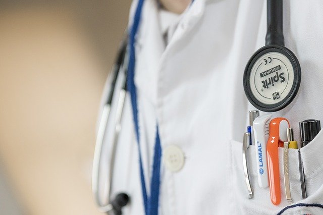
Two hundred and fifty years ago, people lived on average for just forty years. Today, in some industrialized parts of the world, the average lifespan has risen to more than eighty years. Better food and hygiene have helped, but one of the main reasons for this change is the advances made in medicine.
Medicine is the branch of science involved with the prevention, diagnosis, and treatment of disease and damage to the human body. Medical scientists are constantly searching for new ways of treating diseases. Treatments include drugs, radiation therapy, and surgery. Preventive measures, such as vaccinations against infections, are becoming an increasingly important part of modern medicine.
A doctor’s first step with a sick patient is to diagnose the illness. This can be done in various ways: by asking the patient about his or her symptoms, by making a physical examination of the sick person, and by carrying out medical tests if necessary.
Medical treatments may include drugs or surgery. Surgery is the branch of medicine that involves operating to treat the cause of an illness. Today surgery is so advanced that surgeons can sometimes repair or replace organs such as the kidneys and the heart.
Recovery from an illness or an operation may take only a few hours or as long as several weeks. Much depends on the severity of the illness and the impact the treatment has on the body.
Modern medicine makes use of a wide range of technology. The latest developments include body scanners that use a strong magnetic field or ultrasound to produce an image of the interior of the human body. Such equipment has revolutionized medicine.
Перевод
Двести пятьдесят лет назад люди жили в среднем всего сорок лет. Сегодня в некоторых промышленно развитых частях мира средняя продолжительность жизни возросла до более чем восьмидесяти лет. Более качественная еда и гигиена помогли, но одна из главных причин этого изменения ‒ достижения в медицине.
Медицина является отраслью науки, занимающейся профилактикой, диагностикой и лечением заболеваний и повреждений человеческого организма. Ученые-медики постоянно ищут новые способы лечения заболеваний. Лечение включает лекарства, лучевую терапию и хирургию. Профилактические меры, такие как вакцинация против инфекций, становятся все более важной частью современной медицины.
Первый шаг врача к больному пациенту ‒ диагностировать болезнь. Это может быть сделано различными способами: путем опроса пациента о его или ее симптомах, путем физического осмотра больного и, при необходимости, проведения медицинских анализов.
Медицинское лечение может включать лекарства или хирургическое вмешательство. Хирургия ‒ это отрасль медицины, которая включает в себя операцию, чтобы устранить причину болезни. Сегодня хирургия настолько продвинута, что хирурги могут иногда восстанавливать или заменять такие органы, как почки и сердце.
Восстановление после болезни или операции может занять всего несколько часов или даже несколько недель. Многое зависит от тяжести заболевания и воздействия, которое лечение оказывает на организм.
Современная медицина использует широкий спектр технологий. Последние разработки включают в себя сканеры тела, которые используют сильное магнитное поле или ультразвук для получения изображения внутренней части человеческого тела. Такое оборудование произвело революцию в медицине.
Медицина (Medicine) — 5.0 out of
5
based on
1 vote
-
Скачать презентацию (2.05 Мб)
-
261 загрузок -
4.5 оценка
Ваша оценка презентации
Оцените презентацию по шкале от 1 до 5 баллов
- 1
- 2
- 3
- 4
- 5
Комментарии
Добавить свой комментарий
Аннотация к презентации
Презентация для студентов на тему «Medical terms» по иностранным языкам. Состоит из 27 слайдов. Размер файла 2.05 Мб. Каталог презентаций в формате powerpoint. Можно бесплатно скачать материал к себе на компьютер или смотреть его онлайн с анимацией.
-
Формат
pptx (powerpoint)
-
Количество слайдов
27
-
Слова
-
Конспект
Отсутствует
Содержание
-

-
Слайд 2
Скальпель
scalpel [‘skælp(ə)l]
(operating) knife -
Слайд 3
Тампон
pack [pæk]
wick [wɪk]
tampon [‘tæmpɔn]
(surgical) swab [swɔb] -
Слайд 4
ватный тампон:
cotton pellet
cottonwool tampon
марлевый тампон:
gauze [gɔ:z] tampon -
Слайд 5
Электрокардиостимулятор
pacemaker [‘peɪsmeɪkə]
-
Слайд 6
Дефибриллятор
defibrillator [dē’fibrətātər]
-
Слайд 7
Стетоскоп
stethoscope [‘steθəskəup]
-
Слайд 8
Фонендоскоп
phonendoscope
phoneidoscope -
Слайд 9
Тонометр
tonometer[təu’nɔmɪtə]
-
Слайд 10
Ингалятор
inhaler [ɪn’heɪlə]
-
Слайд 11
Вакцина
vaccine [‘væksi:n]
-
Слайд 12
Вата
cotton(wool) [‘kɔt(ə)nwul]
-
Слайд 13
Медицинская груша
spray bulb
-
Слайд 14
Клизма
enema [‘enɪmə]
clysis
clyster [‘klɪstə] -
Слайд 15
Грелка
hot-water bottle
hot-water bag -
Слайд 16
Протез
prosthesis [‘prɔsθɪsɪs]
prosthetic devise -
Слайд 17
Гипс
plaster (of Paris)[‘plɑ:stə]
(dried) gypsum [‘ʤɪpsəm] -
Слайд 18
Жгут
tourniquet [‘tuənɪkeɪ]
garrot -
Слайд 19
Шина
splint [splɪnt]
frame [freɪm]
brace [breɪs] -
Слайд 20
Эмульсия
emulsion [ɪ’mʌlʃ(ə)n]
-
Слайд 21
Рентген
X-ray [‘eks-] photography
radiography[reɪdɪ’ɔgrəfɪ] -
Слайд 22
Рентгенограмма
X-ray [‘eks-] picture
radiograph[‘reɪdɪəugrɑ:f] -
Слайд 23
Флюорография
fluorography[flo͝or’ägrəfē; flôr-]
-
Слайд 24
УЗИ (ультразвуковое исследование)
ultrasound [‘ʌltrəsaund]
US
ultrasonicscanning -
Слайд 25
Томограф
CAT [kæt] (Computerized Axial Tomography) scanner
tomograph -
Слайд 26
Томография
CAT [kæt] (Computerized Axial Tomography) scanning
-
Слайд 27
Костыль
crutch[krʌʧ]
(мн.ч.: pair of crutches)


Посмотреть все слайды
Сообщить об ошибке
Похожие презентации


Спасибо, что оценили презентацию.
Мы будем благодарны если вы поможете сделать сайт лучше и оставите отзыв или предложение по улучшению.
Добавить отзыв о сайте